How the implementation of hydrogen peroxide vapour (HPV) could save you money

On a recent trip to the US, I asked five or six hospitals what their policy was for dealing with packaged medical supplies (syringes, dressings etc) from the rooms of patients on precautions for MDROs. The response was startling: every hospital had a different policy. The policies ranged from 100% disposal of supplies for every discharge through a “toss heavy toss light” approach depending on the perceived risk of the patient to 0% disposal. I scoured international guidelines and, save a few organism-specific guidance documents, there is no direction on this issue in the guidelines.

What actually happens is another issue. Hospitals with a 100% disposal policy knew that staff often could not bring themselves to throw away perfectly good, sometimes fairly valuable items (IV sets and the like) for the small risk that the packaging may be contaminated. Some threw their supplies into “third world bins” to send to underprivileged hospitals, which is great, unless they happen to be contaminated with an MDRO that would survive the journey! Conversely, hospitals with a 0% disposal policy know that sometimes staff threw out the supplies if the patients had a high perceived risk of shedding. As for the “toss heavy toss light” hospitals: who knows what actually happens.

So, is there a real risk associated with contamination of the packaging of these items? A recent study by Johns Hopkins published in the recent ICHE special issue sampled a selection of supplies to quantify the risk, counted the cost of their current policy and found a potential solution in the use of hydrogen peroxide vapour (HPV) for the disinfection of the supplies. The study found that the packaging of 7-9% of supply items was contaminated with MDROs, and that hydrogen peroxide vapor (HPV) was effective for the disinfection of the supply packaging. The cost of supplies discarded from six ICUs amounted to almost $400,000, not including the costs associated with waste disposal. Hence, the practice of disinfecting the packaging of supplies using HPV would generate substantial cost savings.

Can the introduction of 6 copper items into ICU rooms really reduce the rate of HAI by >50%?

The recent article by Salgado et al.in the ICHE special edition is the long-awaited copper study: a multi-centre evaluation of the clinical impact of introducing 6 copper alloy high-touch sites into the rooms of patients on three ICUs. I’ve been looking forward to getting my hands on this study for some time!

copper bed

Patients (n=614 following exclusions) were randomized to intervention ‘copper’ rooms and control ‘non-copper’ rooms in three USA ICUs over an 11 month period. The only difference between the rooms was the presence of six items made of copper alloy, comprising bedrails, overbed tables, IV poles and visitor chair arms in all rooms and the nurse call button, computer mouse, computer palm rest and rim of a touch-screen monitor in other rooms.

Patients admitted to copper rooms were significantly less likely to acquire healthcare-associated infection (HAI) or colonization with MRSA / VRE (Figure).

evidence of copper for hai

Figure: difference in acquisition rate between patients admitted to ‘copper’ or ‘non-copper’ rooms

The authors also make an interesting association between the degree of contamination in patient rooms and the risk of acquisition. However, since sampling was performed weekly regardless of a patient’s infection or colonization status, it is not possible to determine whether this association is causal or simply due to the fact that infected / colonized patients are likely to shed more bacteria into the hospital environment.

The study team should be commended for the careful design and attention to detail in the execution of the study. For example, they performed a daily census of the items in the study to determine exactly who was exposed to copper surfaces, and for how long. This indicated that only half of the patients in ‘copper’ rooms were exposed to all six copper items for the duration of their stay, and 13% of patients in the ‘non-copper’ arm were exposed to some copper items during their stay. It’s important to note that the analysis was performed on an ‘intention to treat’ population, i.e. all patients randomized to the two groups, regardless of which items they were actually exposed to. It would have been interesting to see a sub-analysis on the ‘per protocol’ population (i.e. those patients admitted to ‘copper’ rooms and exposed to all six copper items vs. those patients admitted to ‘non-copper’ rooms and exposed to no copper items). Also, I’m not sure why they chose to analysis % acquisition rather than a comparison of rates between the groups. And then there’s the salary support and grant funding to some authors (not directly related to this study) from the Copper Development Association. Notwithstanding these limitations, it does seem that the introduction of copper high-touch surfaces does reduce the transmission of hospital pathogens.

The scale of the difference is incredible. And I mean just that. It lacks credibility. Many will dismiss the study simply because of the scale of the difference between the groups. So, can the introduction of 6 copper items into ICU rooms really reduce the rate of HAI by >50%?

The degree to which contaminated surfaces contribute to the transmission of pathogens is up for debate. I was asked to give a talk at APIC 2012 on the role of the environment. Prior to the talk, I asked the expert faculty a question: ‘What is your estimation of the % of allC. difficile transmission in hospitals that is mediated, directly or indirectly, by contamination of the inanimate environment?’ The answers ranged from 25-75%, reflecting the uncertainty on this issue.

A recently published study by Johns Hopkins provides some illuminating findings (albeit indirectly). Extrapolating unadjusted acquisition rates from all study cohorts indicates that if rooms had been disinfected using hydrogen peroxide vapour (HPV) after every discharge, the overall rate of acquisition of MDROs would have been halved. Whilst it’s not feasible to disinfect all rooms using HPV at patient discharge, this does add some weight to the scale of the reduction identified in this copper study.

It seems that the introduction of a handful of copper alloy high-touch sites had a profound impact on HAI rates. On reflection, I do believe it possible that the scale of reduction in study is “real”. However, questions remain over the practicality and durability of the widespread adoption of copper alloy surfaces in healthcare.

Article citation:

Salgado et al. Copper surfaces reduce the rate of healthcare-acquired infections in the intensive care unit. Infect Control Hosp Epidemiol 2013;34:479-486.


ICHE special edition on the role of the environment in transmission

Infection Control and Hospital Epidemiology have dedicated their May issue to articles investigating the role of contaminated surfaces in the transmission of pathogens. There’s an awful lot of good stuff here, but this is my take on the key findings of the studies:

  • Drs Weber and Rutala write a thoughtful introduction covering the highlights of the issue.
  • A study from the University of Maryland shows that admission to a room previously occupied by a patient with ESBL-producing Gram-negative bacteria does not increase the risk of acquisition. This is a surprise because this association has been shown for other pathogens including MRSA, VRE, C. difficile and, most interestingly, other Gram-negatives such as A. baumannii and P. aeruginosa. I suspect this difference is explained by the fact that the Enterobacteriaceae are less able to survive on dry hospitals surfaces than the lactose non-fermenting Gram-negatives such as A. baumannii.
  • Research from the Cleveland VA tells a fascinating story of sequential interventions to reduce environmental contamination with C. difficile. The introduction of fluorescent marking with feedback did not eliminate the C. difficile environmental contamination, with 50-60% of cultures remaining contaminated. Similarly, the introduction of a UVC no-touch room disinfection system for terminal disinfection did not solve the problem, with 30-40% of cultures remaining contaminated. Only when daily disinfection was performed by a dedicated team and terminal disinfection was performed by EVS supervisors and/or the infection control team was the problem finally solved and C. difficile could no longer be cultured from surfaces. This study shows firstly how a combination of interventions can be useful, and secondly, the extraordinary lengths required to eliminate C. difficile spores from the environment.
  • An in situ evaluation of a UVC room disinfection device at Duke / University of North Carolina shows that UVC decreases but does not eliminate key pathogens MRSA, VRE and C. difficilefrom the hospital environment.
  • A study from Johns Hopkins shows that the packaging of 7-9% of supply items was contaminated with MDROs, and that hydrogen peroxide vapor (HPV) is effective for the disinfection of the supply packaging. The cost of supplies discarded from six ICUs amounted to almost $400,000, not including the costs associated with waste disposal. Hence, the practice of disinfecting the packaging of supplies using HPV would generate substantial cost savings.
  • The long-awaited copper study is a multi-centre evaluation of the clinical impact of introducing 6 copper alloy high-touch sites into the rooms of patients on three ICUs. Patients were randomized to intervention copper rooms and control non-copper rooms. Patients admitted to copper rooms were significantly less likely to acquire healthcare-associated infection or colonization with MDROs. The authors also make an interesting association between the degree of contamination in patient rooms and the risk of acquisition. However, since sampling was performed weekly regardless of a patient’s infection or colonization status, it is not possible to determine whether this association is causal or simply due to the fact that infected / colonized patients are likely to shed more bacteria into the hospital environment. The scale of the difference is surprising, with a 50% difference between the groups. I am “a believer” in the role of the environment in transmission, but a 50% reduction attributable to 6 copper alloy surfaces does seem rather high. But it does seem that the introduction of copper surfaces does reduce transmission. Questions remain over the practicality and durability of the widespread adoption of copper alloy surfaces in healthcare.
  • Another University of Maryland study with a powerful cluster randomized controlled trial design shows convincingly that enhanced daily cleaning reduces MRSA and MDR A. baumanniicontamination of the gloves and gowns of healthcare personnel when they exit the rooms of patients on precautions with these pathogens. Now, in theory, healthcare personnel should discard the gowns and effectively disinfect their hands. However, since we know that this doesn’t always happen, these reductions are likely to be meaningful.
  • A study shows that N95 filters to their job and capture infectious influenza aerosols. However, in the event of an influenza pandemic, how long will the stockpile of N95 (FFP3) masks last?
  • An extensive microbiology survey from UCLH in London found that the sites closest to the patient were more likely to be contaminated regardless of ward setting. However, in ICUs, sites touched by staff were more likely to be contaminated whereas in gastrointestinal wards with mobile patients, sites touched by patients were more likely to be contaminated.
  • A short review by Carling and Huang explores evolving issues in how to tackle the contaminated healthcare environment.
  • A novel review piece by a team from Georgia, Maryland and Washington DC provides an overview of how evidence-based design can help to prevent and control the transmission of healthcare-associated pathogens.
  • A study from Florida found that 10% of rooms were contaminated with A. baumannii even when the current occupant was not known to be infected or coloinsed. This could be due to unrecognized infection or colonization, survival from a prior room occupant or important by a the patient, a visitor or healthcare personnel.
  • A study from New Haven, Connecticut found that a new activated hydrogen peroxide containing wipe was highly effective for achieving a hygiene standard of <2.5 cfu / cm2, with 75% of sites yielding no growth at all.
  • A new study from the Cleveland VA shows the value of investing time and resource in observing and supervising cleaning practices in hospitals. Direct supervision of cleaning staff was required to achieve optimal results. The concern is what happens when the direct observation becomes routine or stop all together? Will good practice continue?
  • One of the problems with UV radiation for hospital room disinfection is poor reflectivity from some hospital materials, contributing to reduced efficacy out of direct line of sight and influencing cycle times. An innovative study resulting from a collaboration between healthcare experts at the University of North Carolina and chemical engineers at the University of North Dakota found that using paint that reflects UV more effectively reduces cycle times to achieve comparative efficacy for UV room disinfection.
  • A study from South Carolina provides some further microbiological support for the clinical impact associated with copper surfaces, showing that copper alloy bedrails are associated with significantly lower bioburden than plastic bed rails.
  • A useful study from Ireland tells the story of laboratory optimization of sampling methods, which successfully recovered ESBL producing K. pneumonaiaefrom hospital surfaces.
  • A John Hopkins study provided some promising data of a mobile “UV wand” for the disinfection of hospital surfaces. The device a achieve a 1-log reduction of microbial contamination, and may provide a useful adjunctive approach to hospital disinfection.
  • Some Australian data raised some important questions about the reliability of ATP systems. The relative light unit readings for a dilution series of synthetic ATP were compared against an HPLC gold standard for three ATP systems. Substantial variation was noted for all systems, indicating that RLU values are more indicative than absolute.
  • All but one of the studies in this issue have been in the acute healthcare setting. A study from New York bucks the trend, evaluating S. aureus contamination in maximum security prisons. There did not seem to be an obvious association between S. aureus infection and contamination. If anything, the rates of contamination of inmates and their environment was surprisingly, relative to high rates of colonization with MRSA identified in other studies.
  • Yet more research from the Cleveland VA evaluates a novel disinfectant: an electrochemically activated saline solution, also known as ‘superoxidized water’. Surprisingly, the novel disinfectant performed comparably to 10% bleach for the inactivation of C. difficile in vitro, and eliminated C. difficile contamination from hospital surfaces when applied in situ. This agent should be prioritized for further evaluation.
  • Finally, a French study reports a case of catheter-related bloodstream infection related to a preoperative shower with P. aeruginosa contaminated water. Something to think about next time you have a shower.

The quality and importance of the research in this article has impressed me. However, the fact is that some of the basic questions about the role of the environment in transmission and the most cost effective interventions are yet to be answered. But we’re moving in the right direction.

Evidence that iphones are less contaminated than blackberrys?

iphone

Not so long ago, the use of mobile phones in hospitals was banned for both patients and staff. Nowadays, the use of mobile phones by both patients and staff in hospitals has skyrocketed, and is starting to be embraced with the advent of useful clinical apps. However, as the use of mobile phones in hospitals increases, there is a risk that contaminated phones become a new and difficult-to-manage site for contamination and transmission.

I own a blackberry and my wife owns an iphone. We often talk long into the night about the relative benefits of the phones, and the utility of the blackberry keypad compared with the touchscreen of the iphone is my strongest suit. However, a study from UCL in London provides evidence that the keypad of the blackberry is likely to be more contaminated than the touchscreen of the iphone. Sixty-seven phones were sampled, approximately half of which were touchscreen and the other half were keypad phones. Colony counts were significantly higher on keypad phones, which were also more likely to be contaminated with MRSA or VRE. Overall, 13% of the phones grew either MRSA or VRE, highlighting the potential importance of these sites in transmission.

So why the higher counts from the keypad phones? It seems likely that the nooks and crannies on the keypads allow for the buildup of dust and microbial contamination over time, and make the phones more difficult to clean.

It is unacceptable that 13% of mobile phones are contaminated with multidrug-resistant organisms such as MRSA and VRE. Appropriate disinfection policies are urgently required for these personal, mobile reservoirs of contamination.

Article citation:

Pal et al. Keypad mobile phones are associated with a significant increased risk of microbial contamination compared to touch screen phones. J Infect Prevent 2013;14:65-68.

How long does it take to clean a hospital room properly?

Long hours don’t necessarily correlate with productive output. A lifetime’s practice does not necessarily make a champion tennis player. An old boss once told me that “practice doesn’t make perfect; perfect practice makes perfect”. I think there’s something in this that goes some way to explaining the findings of a recent study examining the time taken to clean a hospital room and the thoroughness of cleaning.

You would expect that longer cleaning times would result in more thorough room cleaning. However, the authors used a fluorescent marker to evaluate the thoroughness of cleaning and found no correlation between the length of time cleaning a room and the thoroughness of cleaning.

man_mop

Since this was an assessment of “terminal cleaning” (when the patient was discharged) you would hope that the rates of cleaning for the items in the room would be high. However, the marker was removed from less than half marked sites, and only 5% of monitors were cleaned in the 40 rooms assessed. Disappointingly, there was no correlation between completion of a room a cleaning checklist and removal of the markers.

So, the efficacy of cleaning remains low, even at patient discharge, so it is not surprising that admission to a room previously occupied by a patient with certain multidrug-resistant organisms increases the risk of acquisition!

Article citation: Rupp et al. The time spent cleaning a hospital room does not correlate with the thoroughness of cleaning. Infect Control Hosp Epidemiol 2013;34:100-102.

Micro Blog Spring 2013 update

Dear Readers,

The micro blog has been going well this year, with weekly updates. These are the featured posts:

It would be good to see some more discussion on the blog, so if you have any questions or comments, please don’t be shy! (There was a problem with blocking some email addresses, but that has now been fixed.)

We’ll continue to update the blog with the latest articles weekly. Look out for twitter updates here, and quarterly emails summarizing the top 5 blogs from the last few months.

If you’d like an email every time the blog is updated, just let us know.

Regards

Jon & Saber.

More single rooms = less acquisition. A simple equation?

This isn’t exactly hot off the press (published in 2011) but I’ve only just come across it; it’s a great article and worth revisiting. Many hospitals worldwide suffer a chronic lack of single rooms to place patients requiring contact precautions, but also for patients with other needs such as increased privacy and dignity. There are pros and cons associated with increasing the proportion of single rooms, with a high proportion of single rooms usually requiring a higher staff:patient ratio, and patients in single rooms often having less staff contact and more likely to suffer feelings of isolation. The evidence that an increase in the proportion of single rooms reduces the transmission of healthcare-associated infection has been somewhat equivocal.

This Canadian study evaluated the impact of ‘privatization’ of an ICU. In March 2002, a 24-bed ICU (comprising two 10-bed rooms and four single rooms) was moved to a new 100% single room unit. A 25-bed ICU in a sister hospital (comprising 2, 5, 6 or 8-bed rooms) did not undergo any change in configuration and served as a comparison unit. Importantly, the two units shared the same infection control team, policies and practices. Patients admitted between 2000 and 2005 were studied for the acquisition of a range of pathogens. A unique and useful aspect of the study was to divide microbes into likely endogeneous or exogeneous acquisition. The key result reported was the change in rate ratio of the intervention ICU compared with the comparison ICU before and after the date of privatization of the intervention ICU. This was effectively an estimate of the percentage reduction in the rate in the intervention hospital associated with privatization.

Significant reductions where shown in most pathogens associated with exogenous acquisition, including C. difficile (43%), MRSA (47%) and Acinetobacter spp. (53%), and a substantial but non-significant reduction in Stenotrophomonas maltophilia (52%) (Figure). In addition, a combined analysis of C. difficile, MRSA and VRE also showed a significant reduction of 54%. Significant reductions were also shown for some pathogens in the exogenous/endogenous acquisition group, including Klebsiella spp. (38%). There was no significant change (4%) in the rate of coagulase-negative staphylococci and most other pathogens associated with endogenous acquisition. Perhaps not surprisingly, the authors also reported a reduction in the overall length of stay associated with the intervention.

single room

Figure: Change in the acquisition rate ratio of the intervention vs. comparison ICU before and after before (2001-2002) vs. after (2003-2005) privatisation. (* Not statistically significant.) 

As with all studies, this one is not without criticism. However, there are several aspects that I find particularly convincing. Firstly, whilst there were differences in the configuration of the two ICUs, the inclusion of a comparison unit was an important strength. Secondly, the authors evaluated all available pathogens, rather than focusing on an individual MDRO. Thirdly, and perhaps most convincingly, most pathogens associated with exogeneous infections were affected by the intervention whereas most pathogens associated with endogenous acquisition were not. Additional strengths include several “data-check” sensitivity analyses and an additional model to provide evidence that these were not transient reductions associated with moving to a new, clean unit. Many if not all of these important strengths are lacking from similar studies that have returned a negative result.

The simple equation that more single rooms = less acquisition of pathogens is firmly supported by this study. However, infection rates are not the only factors to be considered when contemplating a move to 100% single rooms. Staffing levels, patients views and up-front costs must be factored into the decision to move towards 100% single rooms.

Article citation:

Teltsch DY, Hanley J, Loo V, Goldberg P, Gursahaney A, Buckeridge DL. Infection acquisition following intensive care unit room privatization. Arch Intern Med 2011;171:32-38.

Carbapenem-Resistant Enterobacteriaceae (CRE) in US hospitals

Enterobacteriaceae are a family of bacteria that commonly cause infections in health-care settings as well as in the community. The family includes more than 70 genera but Escherichia coli, Klebsiella species, and Enterobacter species are the most common in healthcare settings. Until recently, carbapenems have been the treatment of choice for serious infections due to these organisms. However, resistance to these agents has emerged in the Enterobacteriaceae family by various mechanisms and is now a major concern worldwide as infections caused by carbapenem-resistant Enterobacteriaceae (CRE) are difficult to treat and are associated with significant morbidity and mortality.

mmwr

A recent CDC MMWR report used three different surveillance systems to describe the extent of CRE spread among acute-care hospitals in the US as well as the proportion of clinical isolates resistant to carbapenems. They found that while CRE are relatively uncommon, they have spread throughout the US and their rates have increased during the past decade. During the first 6 months of 2012, 4.6% of the 3,918 US acute-care hospitals performing surveillance for either CAUTIs or CLABSIs reported at least once CRE to the National Healthcare Safety Network (NHSN). CRE were more often reported from long-term acute-care hospitals (17.8%) and the percentage of hospitals reporting CRE was highest in the Northeast of the US and among larger and teaching hospitals. Data from NHSN and the Nosocomial Infection Surveillance system (NNIS) showed that the rate of carbapenem resistance among Enterobacteriaceae increased from 1.2% in 2001 to 4.2% in 2011, most of this increase was among Klebsiella species (from 1.6% to 10.4%).

Data from population based surveillance suggest that most (96%) clinical CRE isolates came from cultures collected outside of the hospital from patient with substantial health-care exposure, particularly recent hospitalization (72%). Although nearly all patients with CRE were currently or recently treated in a healthcare setting, these organisms have the potential to spread into the community among healthy individuals.

A combination of infection control strategies applied on national level are needed to control the rise of carbapenem resistance among the Enterobacteriaceae and the spread of CRE. These include active case detection, contact precautions for colonized or infected patients and patient, and staff cohorting as well as strict antibiotic stewardship in all settings. Particular attention should be given to long-term acute-care hospitals which have historically had less developed infection prevention programs. Such coordinated infection prevention and control programs implemented on a national level have been shown to be effective for controlling the rise of CRE, for instance the containment of KPC-producing strains which emerged in 2006 in Israel.

Article citation

Centers for Disease Control and Prevention (CDC): Vital Signs: Carbapenem-Resistant Enterobacteriaceae. MMWR. 2013;62:165-170.

No need to worry about environmental contamination with Enterobacteriaceae…or is there?

It was once thought that only bacterial endospores would survive on dry hospital surfaces for extended periods (measured in days and weeks rather than hours). Microbiological data indicates that a range of vegetative bacteria can survive on dry surfaces for extended periods. Whilst differing testing methods and conditions make comparison of survival times between studies difficult, it is clear that non-fermenting Gram-negative bacteria (such as Acinetobacter baumannii and Pseudomonas aeruginosa) survive considerably longer than the Enterobacteriaceae (such as Klebsiella pneumoniae and Escherichia coli).  However, the Enterobacteriaceae can survive for more than a month on dry surfaces. Indeed, a 2009 laboratory study highlighted substantial strain variation in the survival of K. pneumoniae, with the survival of three strains ranging from a 6-log reduction inside 3 weeks to a 1-log reduction over six weeks.

Several recent studies have evaluated environmental contamination with ESBL-producing Enterobacteriaceae. One French study evaluated surface contamination on five standardized sites surrounding patients infected or colonized with ESBL-producing Klebsiella spp. (n=48) or ESBL-producing E. coli (n=46). Environmental contamination was significantly more likely in the rooms of Klebsiella spp. patients (31% of 48 rooms positive; 6% of 240 sites positive) vs. E. coli patients (4% of 46 rooms positive; 1% of 230 sites sites). Multiple regression identified carriage of ESBL-producing K. pneuomiae as the only independent predictor of ESBL environmental contamination (adjusted odds ratio=10.38, 95% confidence interval = 1.24-228.58). Surprisingly, only 52% of the ESBL-producing isolates were identical to the patients in the room, suggesting survival of ESBL-producing bacteria from prior occupants or importation into the room. Another French study with a similar design identified comparable rates of contamination, and also found that contamination was significantly more likely with K. pneumoniae than with E. coli.

environmental sampling

Environmental contamination with C. difficile spores, VRE and non-fermenting Gram-negative bacteria is now a well-established route of transmission. Whilst the same cannot be said for the Enterobacteriaceae, these studies combined with an Israeli article recently featured on this Micro Blog, show that environmental contamination with Enterobacteriaceae may be more important than previously thought. These findings are particularly important in light of the recent global spread of carbapenemase-producing K. pneumoniae.

Article citations:

Guet-Revillet H, Le Monnier A, Breton N et al. Environmental contamination with extended-spectrum beta-lactamases: is there any difference between Escherichia coli and Klebsiella spp? Am J Infect Control 2012; 40: 845-848.

Gbaguidi-Haore H, Talon D, Hocquet D, Bertrand X. Hospital environmental contamination with Enterobacteriaceae producing extended-spectrum β-lactamase. Am J Infect Cont 2013; Jan 18 [Epub ahead of print].

What does it take to prevent the transmission of C. difficile from environmental surfaces?

Infected patients shed pathogens into the environment, resulting in increased risk of infection for the subsequent occupant of the room by a factor or two or more.1 For example, in one study, patients admitted to rooms previously occupied by patients with C. difficile infection (CDI) were 2.8 times more likely to develop CDI than patients admitted to rooms disinfected using conventional methods. Thus, most agree that more needs to be done to reduce contamination with C. difficile spores in order to interrupt transmission. However, what level of disinfection is required to prevent the transmission of C. difficile?

A consideration of data relating to the infectious dose of C. difficile is a useful first step. In hamster studies, Larson and Borriello showed that only one or two spores of C. difficile were sufficient to initiate infection in clindamycin-treated hamsters.2  This indicates that very low levels of C. difficile spores can initiate infection.

Lawley, et al.3 developed a murine model that provides useful background data on infectious dose. A dose-response relationship was established between the concentration of contamination in the cages and the proportion of healthy mice that developed CDI. All mice became infected when exposed to 100 spores/cm2 and 50 percent of mice became infected when exposed to 5 spores/cm2.  The point at which none of the mice became infected was a concentration of less than one spore per cm2. The mice were exposed to the contaminated cages for one hour. In the healthcare environment, room exposure times are usually measured in days and so these estimates are likely to be conservative. Although translating data from animal models to meaningful clinical practice is difficult, it appears from these animal models that a low concentration of contamination is able to transmit spores to a susceptible host, as is the case with other healthcare-associated pathogens such as norovirus. 4

The low infectious dose of C. difficile is not the only challenge to disinfection. Bacterial endospores can survive on surfaces for many months5 and are resistant to several commonly used disinfectants.6 Patients with CDI result in widespread fecal contamination with C. difficile present in the feces of infected individuals at concentrations in excess of 106 spores per gram.7

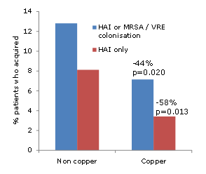
Lawley, et al. examined which disinfectants were able to interrupt the transmission of C. difficile and established a relationship between the level of inactivation of C. difficile spores in vitro and the degree to which transmission was interrupted (figure).

Cdiff_log_reduction

Figure. Correlation between in vitro log reduction and interruption of transmission of C. difficile spores in a murine model.3

The oxidizing agents sodium hypochlorite (bleach) and hydrogen peroxide vapour (HPV) were the only agents tested that achieved a 6-log reduction on C. difficile spores in vitro and completely interrupted the transmission of C. difficile. Notably, both bleach and HPV disinfection can reduce the incidence of CDI in healthcare applications.8-9  Based on these findings, the CDC recommends that surfaces potentially contaminated with C. difficile spores should be disinfected using EPA-registered sporicidal agent (such as bleach) or sterilants.

Recent data highlight the fact that agents with in vitro efficacy may not effectively eradicate hospital pathogens from surfaces due to limitations with achieving adequate distribution and contact time using conventional cleaning methods.1,10 The emergence of ‘no-touch’ automated disinfection methods provide an alternative to reliance on a manual operator for the inactivation of pathogens on surfaces.  HPV is an EPA-registered sterilant that achieves a 6-log reduction on C. difficile spores in vitro, eradicates C. difficile spores from surfaces and reduces the incidence of CDI and successfully mitigates the increased risk from the prior room occupant. 5, 9, 11-13

In summary, given the fact that a small number of C. difficile spores are sufficient to cause CDI in susceptible individuals, disinfectants with an EPA-registered sporicidal claim or sterilants should be used for disinfecting rooms used by patient with CDI. ‘No-touch’ methods, such as HPV, remove reliance on the operator to achieve adequate distribution and contact time and are appropriate for the terminal disinfection of rooms used by patients with CDI.

References:
1. Otter JA, Yezli S, French GL. The role played by contaminated surfaces in the transmission of nosocomial pathogens. Infect Control Hosp Epidem. 2011; 32:687-99.
2. Larson HE, Borriello SP. Quantitative study of antibiotic-induced susceptibility to Clostridium difficile enterocecitis in hamsters. Antimicrob Agents Chemother 1990; 34:1348-53.
3. Lawley TD, Clare S, Deakin LJ, et al. Use of purified Clostridium difficile spores to facilitate evaluation of health care disinfection regimens. Appl Environ Microbiol 2010; 76:6895-900.
4. Yezli S, Otter JA. Minimum infective dose of the major human respiratory and enteric viruses transmitted through food and the environment. Food Environ Microbiol 2011; 3:1-30.
5. Otter JA, French GL. Survival of nosocomial bacteria and spores on surfaces and inactivation by hydrogen peroxide vapor. J Clin Microbiol 2009; 47:205-7.
6. Humphreys PN. Testing standards for sporicides. J Hosp Infect 2011; 77:193-8.
7. Al-Nassir WN, Sethi AK, Nerandzic MM, Bobulsky GS, Jump RL, Donskey CJ. Comparison of clinical and microbiological response to treatment of Clostridium difficile-associated disease with metronidazole and vancomycin. Clin Infect Dis 2008; 47:56-62.
8. Mayfield JL, Leet T, Miller J, Mundy LM. Environmental control to reduce transmission of Clostridium difficile. Clin Infect Dis 2000; 31:995-1000.
9. Boyce JM, Havill NL, Otter JA, et al. Impact of hydrogen peroxide vapor room decontamination on Clostridium difficile environmental contamination and transmission in a healthcare setting. Infect Control Hosp Epidem. 2008; 29:723-9.
10. Manian FA, Griesenauer S, Senkel D, et al. Isolation of Acinetobacter baumannii complex and methicillin-resistant staphylococcus aureus from hospital rooms following terminal cleaning and disinfection: Can we do better? Infect Control Hosp Epidemiol 2011; 32:667-72.
11. Passaretti CL, Otter JA, Reich NG et al. An evaluation of environmental decontamination with hydrogen peroxide vapor for reducing the risk of patient acquisition of multidrug-resistant organisms. Clin Infect Dis 2013; 56: 27-35..
12. Manian FA, Griesnauer S, Bryant A. Implementation of hospital-wide enhanced terminal cleaning of targeted patient rooms and its impact on endemic Clostridium difficile infection rates. Am J Infect Control 2012..
13. Cooper T, O’Leary M, Yezli S, Otter JA. Impact of environmental decontamination using hydrogen peroxide vapour on the incidence of Clostridium difficile infection in one hospital trust. J Hosp Infect 2011; 78:238-40.