Is “community-acquired” CDI real?

A recent high profile US study delved into apparent community-associated CDI cases to evaluate healthcare exposures. The study was large, evaluating almost 1000 cases of community-associated CDI from 8 US states. Only 177 (18%) of the 984 cases had no recent healthcare exposure (Figure 1). Furthermore, healthcare exposure was only evaluated for the 12 weeks prior to the positive specimen, so I would wager that a portion of this 18% acquired their infecting C. difficile in a healthcare facility.  CA-CDIFigure 1. Data demonstrating that most MRSA and CDI presenting on admission to hospital are likely to have been acquired in a healthcare facility.  

So, it seems that the majority of these cases are more likely to be community-onset, healthcare-acquired CDI, rather than community-acquired CDI. I feel like we’ve been here before. In the 1990s before the emergence of distinct strains of CA-MRSA, MRSA presenting at hospital admission was commonly termed ‘community-associated’ or, worse, ‘community-acquired’ when really it was MRSA that had been acquired in hospital during a previous stay (Figure 1). The situation has now changed since distinct MRSA clones have emerged that have the capacity to cause infection outside the healthcare environment.

Turning our attention to the UK, the mandatory report scheme classifies cases of CDI as ‘Trust-apportioned’ if the specimens is collected from patients who have been in hospital for four or more days (Figure 2). It is tempting to speculate that the cases of CDI that are non Trust-apportioned are CA-CDI. However, the definition for ‘Trust-apportioned’ does not account for previous healthcare contact, and the rate of Trust-apportioned and non-Trust-apportioned cases tracks so closely that, once again, these are likely to be healthcare-acquired CDI presenting on admission.

Slide1Figure 2. Number of cases of CDI in England through the mandatory reporting scheme, 2004-2013.

The epidemiology of C. difficile is fundamentally different to MRSA, in that healthy neonates typically have a high rate of C. difficile colonization. Thus, there is a ready reservoir for a low rate of genuinely community-acquired CDI. However, it seems to me that most “CA-CDI” reported thus are likely to be acquired in a healthcare facility and I have not seen any data to convince me that community-acquired CDI is increasing.

Article citation: Chitnis et al. Epidemiology of Community-Associated Clostridium difficile Infection, 2009 Through 2011. JAMA Intern Med 2013;173:1359-67.

Is there a causal relationship between contamination burden and transmission risk?

contamination v transmission There’s an age-old problem in science: how do you prove a causal relationship between variables that correlate? Proving that the variables are correlated is the easy part; it’s more difficult to disentangle cause from effect. This can be seen in several studies that identify a correlation between environmental burden and the number of patients that are infected or colonized with pathogens.DentonFigure 1. Correlation between the number of patients infected with Acinetobacter spp. and the number of positive Acinetobacter spp. environmental cultures per calendar month during an outbreak on a neurosurgical ICU.1

SalgadoFigure 2. Correlation between microbial burden and the number of patients who acquired an HAI in ICUs.2

WhiteFigure 3. Correlation between the number of hygiene failures and the number of patients who acquired an infection on a surgical intensive care unit each week.3

So can we conclude that the higher burden of contamination resulted in an increased risk of acquisition? Or is it that more patients were infected or colonized with pathogens, which resulted in more environmental shedding? From these studies, you can’t be sure.

If you were seeking to prove the role of a gene in a process, you’d knock out the gene and demonstrate that the process stopped or changed. So, the only way to disentangle cause and effect in contamination and transmission is to perform an intervention to reduce environmental contamination and show that this correlates with reduced transmission. While the Salgado study evaluated an intervention, the data correlating contamination burden with HAIs was not stratified by the intervention, which would have been one way to assess likely causation.2

There is some further in vitro and epidemiological data supporting that the degree of transmission may be proportional to the environmental burden. An in vitro mouse model established a ‘dose-response’ relationship between the degree of contamination with C. difficile spores and the development of CDI.4 Furthermore, this model showed that disinfectants that achieved a greater log reduction of C. difficile spores were more able to interrupt transmission.

Also, one of the studies demonstrating that admission to a room previously occupied by a patient with VRE increases the chances of VRE acquisition identified something amounting to a ‘dose response’.5 The greatest increased risk was for patients admitted to a room with an environmental culture positive for VRE, and being admitted to a room where the immediate prior room occupant was colonized with VRE carried a greater increased risk than being admitted to a room where any patient in the 2 weeks prior to admission was VRE colonized (Figure 4).

DreesFigure 4. How the increased risk of acquiring VRE from the prior room occupant changes due to patient and environmental factors.5

Is there a causal relationship between contamination burden and transmission risk? On balance, the answer seems to be yes, though it would be useful to have a solid intervention study to prove that an increasing environmental burden causes an incrementally increase in transmission risk.

Article citations:

  1. Denton M, Wilcox MH, Parnell P et al. Role of environmental cleaning in controlling an outbreak of Acinetobacter baumannii on a neurosurgical intensive care unit. J Hosp Infect 2004; 56: 106-110.
  2. Salgado CD, Sepkowitz KA, John JF et al. Copper surfaces reduce the rate of healthcare-acquired infections in the intensive care unit. Infect Control Hosp Epidemiol 2013; 34: 479-486.
  3. White LF, Dancer SJ, Robertson C, McDonald J. Are hygiene standards useful in assessing infection risk? Am J Infect Control 2008; 36: 381-384.
  4. Lawley TD, Clare S, Deakin LJ et al. Use of purified Clostridium difficile spores to facilitate evaluation of health care disinfection regimens. Appl Environ Microbiol 2010; 76: 6895-6900.
  5. Drees M, Snydman D, Schmid C et al. Prior environmental contamination increases the risk of acquisition of vancomycin-resistant enterococci. Clin Infect Dis 2008; 46: 678-685.

The pitfalls of PCR for detecting pathogens on surfaces

PCR has proven an invaluable tool for the rapid diagnosis of a range of pathogens, including MRSA and C. difficile. Several studies have evaluated the potential use of PCR for the detection of pathogens on surfaces and have identified some issues that, frankly, seem pretty terminal for this application using currently available commercial PCR kits.

A study from Cleveland evaluated the use of a commercial RT-PCR test for detecting C. difficile on hospital surfaces. Three composite sites were sampled in 22 patient rooms, 41% of which housed a patient with CDI with the remaining 59% sampled after terminal cleaning and disinfection. Two swabs and a gauze were collected from each site; one swab was cultured directly onto selective agar and the other was tested using PCR. The gauze was cultured using broth enrichment. C. difficile that grew on the selective agar were tested for toxin production and only toxigenic C. difficile were included.

Overall, 23 (35%) of the 66 sites grew toxigenic C. difficile and only 4 of these were detected using the standard RT-PCR assay (sensitivity 17%, specificity 100%). The sensitivity of RT-PCR in rooms that had been cleaned and disinfected was even worse (10%). Increasing the CT threshold of the assay (making it less stringent) improved the overall sensitivity to 52% and did not affect the specificity.

The study has several important limitations. The RT-PCR assay detected only the Toxin B gene, whereas the toxigenic culture methodology would detect both Toxin A and B producers. More importantly, there was a crucial difference in sampling methodology: the gauzes used for broth enrichment culture had a 50% higher positivity rate than the swabs (in line with other findings), but only swabs were tested by both PCR and culture. Thus, if the gauzes are a more effective sampling device, this would make the RT-PCR methodology seems worse than it is. I would have liked to have seen the sensitivity of the RT-PCR assay for detecting C. difficile cultured from the swabs only, but I could not derive this from the data in the paper.

An older study from New Haven, Connecticut provides a contrasting view of the use of PCR to detect pathogens from surfaces. Here, 10 standardized sites were sampled in the rooms of 10 patients infected or colonized with MRSA, and 5 rooms of patients not known to be infected or colonized with MRSA. Swabs were directly plated onto selective agar for MRSA, then DNA was extracted from the swabs before a broth enrichment procedure using the same swabs. In this study, 40 (27%) of the 150 surfaces were positive by culture, but 90 (60%) were positive by PCR (sensitivity 93%, specificity 51%).

Deshpande 2013

Figure 1. Contrasting sensitivity and specificity when using PCR to detect C. difficile and MRSA on hospital surfaces.

It seems then that the sensitivity of PCR is too low for the environmental detection of C. difficile but the specificity is too low MRSA (figure 1). How could this be? Assuming that this is not due to experimental differences between the studies, it could be that the standard extraction procedure used for the C. difficile assay was not robust enough to liberate DNA from the mature environmental spores, resulting in low sensitivity. Conversely, the PCR assay was detecting DNA from dead MRSA on surfaces, resulting in low specificity.

So, in summary, the MRSA assay was too sensitive and the C. difficile assay was not sensitive enough! While the use of these “off the shelf” commercial assays doesn’t seem to be useful for detecting pathogens on surfaces, there may be hope for a PCR assay tailored specifically for an environmental application.

Article citations:

Deshpande A, Kundrapu S, Sunkesula VC, Cadnum JL, Fertelli D, Donskey CJ. Evaluation of a commercial real-time polymerase chain reaction assay for detection of environmental contamination with Clostridium difficile. J Hosp Infect 2013;85:76-78.

Otter JA, Havill NL, Boyce JM. Evaluation of real-time polymerase chain reaction for the detection of methicillin-resistant Staphylococcus aureus on environmental surfaces. Infect Control Hosp Epidemiol 2007;28:1003-1005.

Is it time to turn to ‘no-touch’ automated room disinfection?

I gave a webinar for 3M yesterday entitled ‘Is it time to turn to ‘no-touch’ automated room disinfection (NTD)?’ It was based broadly on a recent Journal of Hospital Infection review article, and you can access the slides here.

The webinar covered:

  • The key data supporting the need for improved hospital disinfection, particularly ‘terminal disinfection’ when patients are discharged.
  • The strengths and limitations of conventional disinfection methods, particularly in terms of reliance on the operator to ensure adequate formulation, distribution and contact time of the active agent.
  • The potential benefits of introducing automation into the room disinfection process.
  • Coverage of the advantages and disadvantages of the various “no-touch” automated room disinfection systems currently available.
  • Scenarios in which NTD systems may be warranted.

To summarize the rationale for using an NTD system: enhanced conventional methods are able to eliminate pathogens from surfaces, but the inherent reliance on a human operator to ensure adequate formulation, distribution and contact time of the active agent introduces variability into the process. NTD systems remove or reduce reliance on the operator for delivering hospital disinfection. However, they do not obviate the need for cleaning, so they are designed to augment rather than replace conventional methods.

So when to consider an NTD system? The flow chart below (Figure 1) shows a decision tree for which cleaning and disinfection approach to take. Given their practical limitations, NTD systems are best suited to disinfection of a room after a patient colonized or infected with a pathogen has been discharged to protect the incoming patient from acquiring the pathogen left behind by the prior room occupant. A recent study of a hydrogen peroxide vapor (HPV) NTD system shows that patients admitted to rooms disinfected using HPV were 64% less likely to acquire any multidrug-resistant organism (MDRO) than patients admitted to rooms disinfected using standard methods when the prior room occupant had an MDRO.Flow chartFigure 1. A disinfection decision diagram for when to consider an NTD system. a) Key pathogens associated with contamination of the environment include C. difficile, VRE, MRSA, A. baumannii, P. aeruginosa and norovirus. b) All NTD systems are applied after a cleaning step to ensure that surfaces are free from visible contamination, which is unacceptable to subsequent patients and will reduce the efficacy of the NTD disinfection. c) There is limited equivocal evidence that enhanced cleaning / disinfection in a low-risk general ward setting can reduce the spread of pathogens.

Ok, so you’ve decided that you want to use an NTD system. Which one to choose? Every conference I go too seems to have more and more NTD systems on show, all with bold and often conflicting claims. There are essentially four classes of NTD system that are commonly used in hospitals:

  • Hydrogen peroxide vapor (HPV)
  • Aerosolized hydrogen peroxide (aHP)
  • Ultraviolet C (UVC)
  • Pulsed-xenon UV (PX-UV)

I asked the audience which, if any, NTD system had been used in their hospital (Figure 2). 90% of the predominantly US based audience had not used an NTD system at all, which was a surprise. In the hospitals that had used an NTD system, there was a fairly even split between HPV and the UV systems.Which systemFigure 2. Has your hospital used an NTD system and if so, which one?

Each of these systems have advantages and disadvantages, which I have tried to summarize in the following table by ranking the systems in the key categories. The hydrogen peroxide systems tend to have higher efficacy and better distribution than the UV systems. But the UV systems are faster and easier to use. Thus, there is a trade-off between efficacy / distribution and cycle time / ease of use when deciding which NTD system would be more appropriate.Comparision table Table: Comparing the key features of the four commonly used NTD systems.

In order to illustrate the challenges in choosing a) whether to use and NTD system and b) which to use, I presented the audience with three scenarios. In scenario 1, below, I was expecting most people to select ‘conventional methods’ or one of the UV systems, which have both been shown to reduce the burden of contamination without reliably eliminating pathogens. The sheer number of patients with MRSA colonization transferred or discharged from general medical wards means that the additional time for HPV may not be warranted.Scenario 1Scenario 1. What do you do when a patient who was colonized with MRSA has been discharged from a room on a general medical ward?

Scenario 2 is an occasion where you want to be sure that residual contamination has been dealt with so that the incoming susceptible ICU patient will not acquire the virtually untreatable carbapenem-resistant A. baumannii. Therefore, HPV, which is associated with the elimination of pathogens from surfaces, is a rational choice.   Scenario 2Scenario 2: What do you do when a patient who had an infection with carbapenem-resistant A. baumannii has been discharged from an ICU room?

Scenario 3 is more tricky. While the likelihood of C. difficile spore contamination argues for the higher efficacy of the hydrogen peroxide systems, the number of transfers or discharges of patients with C. difficile on a surgical unit may be high, which argues for the lesser efficacy but faster cycles from the UV systems. The majority of the audience selected HPV in this scenario, considering that the combined risk of the pathogen and specialty required the elimination of C. difficile spores from the room prior to the admission of the next patient.    Scenario 3Scenario 3: What would you do when a patient who had C. difficile infection has been discharged from a room on a surgical unit?

To summarize, the use of an NTD system to augment terminal disinfection is warranted in some circumstances. The choice of NTD system will depend on a number of factors, including efficacy, distribution, ease of use, cycle time and cost. The features of the various NTD systems make them best suited to different applications, dictated by the clinical setting and the environmental-pathogenic characteristic of the target pathogen. So, is it time to turn to NTD systems? 52% of the audience voted ‘yes’ at the start of the webinar; 74% voted ‘yes’ at the end!Initial finalFigure 3: Is it time to turn to ‘no-touch’ automated room disinfection? The audience were asked this question at the start and the end of the webinar, indicating a swing towards the affirmative!

Article citation: Otter JA, Yezli S, Perl TM, Barbut F, French GL. Is there a role for “no-touch” automated room disinfection systems in infection prevention and control? J Hosp Infect 2013;83:1-13.

The effect of closing and cleaning wards on infection rates

Not so long ago, the UK Government ordered a national ‘deep clean’. This prompted a fair amount of debate among experts and the public. If the NHS needed a spring clean, then does that mean that it was dirty in the first place? Perhaps. There does not seem to have been a formal evaluation of impact, but there is some rationale for closing and cleaning wards. For example, this paper from the early 1970s evaluated the impact of closing and cleaning five wards in London.

The five wards (four surgical and one medical) had an outbreak of MRSA (termed ‘cloxacillin-resistant S. aureus’). Rates of infection (termed ‘sepsis’) were monitored on the study wards before and after closing and cleaning. Wards were closed to admissions and emptied of patients. All fabrics were sent for laundering and all left over supplies were discarded. Cleaning comprised washing floors, walls and all other surfaces with hot water containing detergent; bed frames and furniture were also washed. The length of time that all this cleaning too is not specified, but I suspect it took place over several days. Crucially, staff and patients were screened for carriage of epidemic strains of S. aureus; colonised patients were not re-admitted after ward cleaning where possible.

The charts below show the impact on all infections (Figure 1), all S. aureus infection (Figure 2) and MRSA infection (Figure 3). Infection rates were compared 3 months before vs. 3 months after cleaning on Wards 1-3 and 6 months before vs. 6 months after on Wards 4 and 5. As you can see, the impact was pretty dramatic.

Noone Fig 1

Figure 1. Total infection rate (proportion of admissions infected) on the five wards before vs. after ward closing and cleaning.

Noone Fig 2

Figure 2. S. aureus infection rate (proportion of admissions infected) on the five wards before vs. after ward closing and cleaning.

Noone Fig 3

Figure 3. MRSA infection rate (proportion of admissions infected) on the five wards before vs. after ward closing and cleaning.

The poor reduction in total infection rate on Ward 1, a gynecological ward, (Figure 1) is largely due to high Gram-negative infection rates before and after cleaning, most likely explained by endogenous urinary tract infections. Reductions in total infection rate and S. aureus infection rate appeared to be less on Wards 4 and 5, which could be influenced by the fact that rates were compared for 6 months pre and post ward closing and cleaning rather than 3 months on Wards 1-3. The impact of a one off environmental intervention is likely to diminish over time. It’s also interesting to note that the MRSA infections identified on Ward 5, a general surgical ward, after cleaning were due to a different strain of MRSA (determined by phage typing and antibiogram) than before cleaning. This new strain matched the outbreak strain from Ward 2. Two of the patients on Ward 5 who became infected with this strain were operated on in the same theatre as the infected patients from Ward 2 within two weeks of one another. Four other patients (on different wards) also appeared to acquire the strain in the same operating theatre.

The study has several important limitations. It is not possible to be certain whether active screening and isolation or ward closing and cleaning were responsible for the reduction in infection rates; it was probably combined impact. The study design lacked the rigor of more modern investigations: infection rates were not expressed in terms of patient-days and infection rates were compared for different time periods making direct comparison of the impact across the five wards difficult. Also, no environmental sampling was conducted to demonstrate the efficacy of the cleaning procedure (both initially and in terms of recontamination).

Notwithstanding these limitations, the study provides evidence that ward closing and cleaning combined with active screening and selective readmission resulted in a dramatic reduction in the rate of nosocomial infection on five study wards. The impact appeared to be most pronounced in the first three months, which is consistent with a reduction in environmental contamination. Outbreaks of MRSA were eradicated by closing and cleaning on all five study wards. However, there was evidence of new nosocomial transmission following the re-admission of infected patients. Finally there was some interesting circumstantial evidence of transmission within operating theatres.

Article citation: Noone P, Griffiths RJ. The effect of sepsis rates of closing and cleaning hospital wards. J Clin Pathol 1971;24:721-725.

Prevalence survey illustrates the difference between CRE and CPE

I recently posted an article on the difference between CPE and CRE, which is neatly illustrated by a prevalence survey from Alder Hey Children’s Hospital in Liverpool. In case you didn’t read my CPE/CRE blog (shame on you), here’s a reminder of the difference between the two:

Carbapenem-resistant Enterobacteriaceae (CRE) – Enterobacteriaceae that are resistant to carbapenems by any mechanism, including the production of an acquired carbapenemase or the production of an ESBL or AmpC combined with porin loss.

Carbapenemase-producing Enterobacteriaceae (CPE) – Enterobacteriaceae that are resistant to carbapenems by means of an acquired carbapenemase.

At Alder Hey, a large children’s hospital in Liverpool, a prevalence survey was performed between September 2011 and August 2012. All clinical and screening specimens were included; rectal screens were collected on admission and weekly from all patients in the ICU and HDU. 24 patients with CRE were identified, five (21%) from clinical specimens and 19 (79%) from rectal screens. The prevalence of CRE in rectal screens was 4.5% (19/421). Four of the 19 patients identified by screening specimens only went on to develop an infection, so 9 (38%) of patients ended up with a CRE cultured from a clinical specimen.

The majority (71%) of the 24 isolates were resistant to carbapenems by AmpC or ESBL combined with impermeability; seven (29%) were CPE, 4 with NDM and three with KPC (Figure). Typing indicated that the 3/4 NDM producing Klebsiella pneumoniae isolates were clonal, and they were clustered in space and time, which may indicate a small outbreak.

CRE alder hey Figure. Composition of CRE at Alder Hey Children’s Hospital. 

Carbapenem-resistance due to the production of an ESBL or AmpC combined with porin loss may lead to treatment failure, but it is often unstable and may impose a fitness cost, meaning that these strains rarely spread. Hence, carbapenem resistance conferred by an acquired carbapenemases is the key problem. This study helps to define the prevalence of CRE (and, more importantly CPE) in the population. We are not given a denominator for the clinical specimens, so the prevalence of CRE amongst clinical specimens cannot be calculated. However, the fact that around 5% of patients admitted to ICU / HDU were carrying CRE is a concern, although the prevalence of CPE on the rectal screens was lower at 1.7% (7/421).

Currently, the prevalence and epidemiology of CRE and CPE is poorly defined in the UK so this useful prevalence survey from Alder Hey is welcome. However, we urgently need more research from other hospitals to scale the CRE problem.

Article citation: Drew et al. Emergence of carbapenem-resistant Enterobacteriaceae in a UK paediatric hospital. J Hosp Infect 2013;84:300-304.

Look out: resistance outbreaks about!

We are all familiar with the idea of outbreaks. A noteworthy pathogen rears its ugly head leaving a trail of destruction in its wake (as in ‘Contagion’, or before that, ‘Outbreak’). (I credit ‘Outbreak’ with getting me into microbiology and epidemiology as an impressionable 15 year old by the way.) Or more commonly in hospitals, a ward experiences an increased incidence of a particularly resistant or virulent clone. But a recent study from some colleagues at the Centre for Clinical Infection and Diagnostics Research at St. Thomas’ Hospital in London turns the idea of ‘outbreak’ on its head by identifying surprisingly common outbreaks of resistance to a particular antibiotic across different species.

The horizontal transfer of resistance genes is generally considered to be a rare event relative to horizontal, clonal transmission of an outbreak pathogen (see Figure below). But the findings of this study suggest more promiscuous spread of resistance genes than you may expect.

ICU resistance

Figure: Horizontal transfer of resistance genes is generally considered to be the least common cause of ICU resistance.  

The team used some outbreak scanning software to interrogate laboratory reports from two ICUs between 2002 and 2009. Analysis of the large dataset, comprising almost 90,000 patient days, found that outbreaks occurred for two thirds of the 26 ‘species-groups’ studied. Only three of these were recognized at the time. Thirty-nine outbreaks of resistance were detected, the majority of which (87%) did not coincide with an increase in a particular ‘species-group’, supporting the fact that these were due to horizontal gene transfer between species.

The clustering of individual species into ‘species-groups’ is somewhat problematic, and may serve to over-emphasize the number of outbreaks that occurred. Quite a number of the outbreaks of the same ‘species-group’ and of resistance were very small – with 2 cases over a day or two. Also, clustering of the same species does not necessarily mean clonal transmission has occurred – you’d need to do molecular typing to prove that. Similarly, clustering of resistance across species to the same antibiotic does not necessarily mean horizontal gene transfer has occurred; multiple mechanisms could be involved. Notwithstanding these limitations, this is an important study and has changed the way that I think about hospital outbreaks.

Infection control interventions implemented to control recognized outbreaks on the ICU appeared to reduce the overall number of outbreaks of the same ‘species-group’, but did not affect the number of resistance outbreaks. So, it seems that different measures are necessary to control outbreaks of resistance. Perhaps the best weapon we have to combat outbreaks of resistance is to restrict our use of antibiotics. If we can reduce the selective pressure driving resistance, we should see less clonal outbreaks of resistant bacteria and less resistance outbreaks across species.

Article citation: Vlek AL, Cooper BS, Kypraios T, Cox A, Edgeworth JD, Auguet OT. Clustering of antimicrobial resistance outbreaks across bacterial species in the intensive care unit. Clin Infect Dis 2013; 57: 65-76.

Do you know your CRO from your CPO from your CRE from your CPE?

Carbapenems are a class of beta-lactam antibiotic with a broad spectrum of activity against Gram-positive and Gram-negative bacteria. Whilst carbapenems are used for the treatment of Gram-positive infections, the emergence of Gram-negative bacteria with resistance to the carbapenem antibiotics is a health issue that has prompted unusually dramatic health warnings from the US CDC, Public Health England (PHE) and the European CDC (ECDC). However, the various acronyms employed to describe the subtleties of the problem are a minefield for the uninitiated:

Carbapenem-resistant organism (CRO) – Gram-negative bacteria* including the Enterobacteriaceae (such as Klebsiella pneumoniae and Escherichia coli) and non-fermenters (such as Acinetobacter baumannii, Pseudomonas aeroginosa and Stenotrophomonas maltophilia) that are resistant to carbapenems by any mechanism. The non-fermenters can be inherently resistant to carbapenems, or they can acquire carbapenemases (typically KPC, VIM, NDM and OXA-48 types). Enterobacteriaceae do not have inherent resistance but may be resistant to carbapenems through the production of an acquired carbapenemase or the production of an ESBL or AmpC combined with porin loss.

Carbapenemase-producing organism (CPO) – Enterobacteriaceae and non-fermeters that are resistant to carbapenems by means of an acquired carbapenemase.

Carbapenem-resistant Enterobacteriaceae (CRE) – Enterobacteriaceae that are resistant to carbapenems by any mechanism, including the production of an acquired carbapenemase or the production of an ESBL or AmpC combined with porin loss.

Carbapenemase-producing Enterobacteriaceae (CPE) – Enterobacteriaceae that are resistant to carbapenems by means of an acquired carbapenemase.

The image below tries to graphically represent the relative size of these groups (not to scale!), and the table provides a summary of their distinguishing features:

CRE etc

CRE table

“O” or “E”

The US CDC and European CDC seem to favour ‘CRE’ as a generic term for this problem, whereas the PHE in the UK seems to favour ‘CRO’. Clearly, each term has a defined meaning and context will determine which is technically correct. But which is most useful as a generic term?

The epidemiology of the non-fermenters and the Enterobacteriaceae is different, with the non-fermenters tending only to cause problems in very sick patients, usually in critical care settings. Meanwhile, the Enterobacteriaceae are more able to cause infections in a wider range of patients both inside and outside the hospital. Hence, the emergence of carbapenem resistance is more concerning in the Enterobacteriaceae, as demonstrated by rapid national and international spread of KPC-producing K. pneumoniae.

“R” or “P”

Amongst the Enterobacteriaceae, whilst carbapenem-resistance due to the production of an ESBL or AmpC combined with porin loss may lead to treatment failure, it is often unstable and may impose a fitness cost, meaning that these strains rarely spread. Hence, carbapenem resistance conferred by an acquired carbapenemases is the key problem.

So, for me, CPE would be the most suitable generic term for this emerging problem. However, since the US CDC and the ECDC seem to have gone with CRE, and the vast majority of CRE will be CPE, let’s go with CRE shall we?


[* Whilst Gram-positive bacteria that are resistant to carbapenems (such as MRSA) could be described as ‘CROs’, these terms are reserved to describe Gram-negative bacteria.]

This is what happens when norovirus “sprays” from a toddler

baby changeAn outbreak report in the Journal of Infectious Diseases tells the fascinating story of a norovirus outbreak in a car (auto*) dealership in Oregon that was initially thought to be foodborne, but was eventually traced to contaminated surfaces on a baby changing table (diaper changing station*) in a public toilet (restroom*). The outbreak had a startlingly high attack rate, affecting 75% of 16 employees who attended a team meeting. A thorough investigation of the restaurant that provided the sandwiches for lunch turned out to be a blind alley following the recollection of a staff member of a toddler with “spraying” diarrhoea using the baby changing table in the public toilet of the dealership. The (generous) mother left the mess for the staff member to clean up, which was accomplished using, wait for it, dry paper towels.

The environmental investigation included samples from the baby changing table in the dealership and some ‘control’ samples from 14 baby changing tables in public toilets throughout the state. Norovirus of the same genotype as the outbreak strain was identified from the baby changing table in the car dealership, but norovirus was not identified from the control baby changing tables.

Some limitations of the outbreak include the fact that it is difficult to disentangle the relative importance of the environmental reservoir and secondary transmission via contaminated food. The PCR method used for environmental sampling does not assure that the norovirus RNA identified on the baby changing table was viable. Also, the environmental norovirus isolates could not be sequenced meaning that they could not be sequence-matched with the patient isolates.

Perhaps the most shocking part of the story is that the image of visible soiling on the baby changing table (after two rounds of cleaning) was consistently viewed on baby changing tables in public toilets. Or perhaps it’s even more shocking that only 3 of the 12 affected individuals actually took time off work. As a “survivor” of a norovirus outbreak that swept through the Otter household in 2012, I can vouch for the fact that a) you need to take time off work and b) you ought to take time off work!

One important discussion point was the finding of Dr Carling’s group that baby changing tables were least likely to be cleaned on cruise ships, a setting in which persistent norovirus outbreaks are common. This outbreak report and Dr Carling’s earlier work highlight an important deficiency in how to clean and disinfect baby changing tables in public toilets. As a frequent user over the past 14 months, I can vouch for the fact that, much like a hospital bed, turnover is high which pressurizes effective terminal disinfection! I agree with the authors that disinfection with a chlorine-containing disinfectant would be ideal, but question whether this is feasible in practice.

There’s surprisingly little data supporting the role of environmental contamination in the transmission of norovirus. I’m persuaded by the various outbreaks affecting separate cohorts of patients / staff on cruise ships or aeroplanes, but this outbreak is even more compelling due to the environmental findings. Quantifying the role of the environment in the transmission of norovirus is difficult to study because it always occurs in outbreaks (hence difficult to perform a controlled study). But I’d be interested to see whether the “prior room occupancy” concept that has been established for other environmentally-associated pathogens holds true for norovirus.

Article citation: Repp KK, Hostetler TP, Keene WE. A norovirus outbreak related to contaminated surfaces. J Infect Dis 2013;208:295-298.

Also, take a look at Dr Repp’s blog.

* = for my American readers! Current data indicates that around 40% of the readers of this blog are US based, 40% are UK based and 20% are rest of world!

ICPIC 2013 Conference Report

icpic2013_banner

My head is full and my wallet is light following an enjoyable week in Geneva for the 2nd ICPIC conference (25th – 28th June 2013). I missed out on the inaugural ICPIC in 2011, so I was pleased to make the 2nd ICPIC. I was a little concerned that it would be a “hand hygiene fest”, but the programme had a good balance between hand hygiene and other areas of infection prevention and control. The conference had more than 900 delegates from 89 nations with 97 oral presentations and 427 posters. Congratulations to the organizing committee for arranging an interesting and stimulating conference. The conference abstracts are freely available in an ARIC supplement and the slides can be purchased by contacting the conference organizers.

Sadly I was unable to clone myself and attend every session, but this will give you a flavour…

TUES 25TH JUNE – OPENING SESSION

The opening session focused on viruses with pandemic potential, with an impressive introduction from some Genève luminaries and a personal video from Dr Margaret Chan (WHO Director General).

David Heymann – The legacy of SARS

Dr Heyman presented an engaging first-hand overview of the early reports of SARS, illustrating that it was predominately a healthcare-associated infection. The legacy of SARS according to David Heymann:

  1. Improved global surveillance, for example, the advent of new surveillance methods and networks such as Google outbreak software.
  2. “Research ready” capacity. The emergence of new microbial threats promotes the requirement for a research network ready and waiting to swing into action, such as the Oxford University Institute for Emerging Infections.
  3. Economic impact. SARS cost Hong Kong 4% of GDP.
  4. Understanding animal reservoirs. HIV, SARS, Avian Influenza, MERS-CoV (?) and most other pathogens with pandemic potential have emerged from animal reservoirs.

Wing Hong Seto – Infection control before and after SARS

Dr Seto’s thesis was that the spread of SARS in hospitals was due to poor basic infection control practice, evidenced by his own institution’s low rate of staff infection stemming from his enthusiastic education and awareness campaign. Put another way, sloppy infection control cost healthcare professional’s their lives. Dr Chan stated earlier that “the thermometer won the war against SARS”, which resonated with Dr Seto’s “getting the basics right” message. The experience of SARS has resulted in an increase in focus on infection control in Asia region, resulting in more investment, higher infection control staff : bed ratios and centralized expert committees. However, there has been some overreaction too, with very expensive structural changes to hospitals without proper consultation. Dr Seto’s  enthusiasm truly lit up the stage.

Laurent Kaiser – influenza, coronavirus and emerging viruses: can we predict the unexpected?

A detailed overview of the genetic basis of these pandemic viruses that are associated with respiratory disease, aerosol / droplet spread, seasonal patterns, an RNA genome and rapidly emerging strains. The most interesting part of the talk for me was a consideration of how long it takes to “humanize” an animal virus. Whilst it’s difficult to be sure, it seems that it takes decades (or perhaps even centauries) for the necessary amount of recombination and mutation to occur for a virus to jump to humans and spread efficiently. For example, the H1N1 virus seems to be a mosaic of three viruses that may trace its origins to the 1918 pandemic. In this age of almost instant phylogeny, it may not be long before we can start watching viruses evolve in real time.

Keiji Fukuda – Latest news from MERS-CoV and H7N9

Dr Fukuda (WHO) gave a brief historical view of HIV, SARS, H5N1 and H1N1 to illustrate how a pandemic response looks in relation to the two current threats: H7N9 and MERS-CoV. A relatively small number of N7N9 (132) and MERS-CoV (70) cases have been reported so far, but their high mortality rates are alarming. Interesting unanswered questions:

  1. Are we at a suitable level of readiness to respond?
  2. How should we name pandemic viruses?
  3. How do we handle intellectual property that emerges during pandemic response?

Robert Wachter – Embedding infection prevention into medical training and the patient safety agenda

Dr Wachter explained that the patient safety movement began with infection control, but is now predominant in the US. Infection control should seek to embed itself within the core values of patient safety: quality and value. Dr Wachter explained some seminal moments in the development of the patient safety movement: the realization that medical errors were killing the equivalent of a jumbo jet of Americans each day and that zero is possible. Perhaps it would be helpful to see non-compliance with hand hygiene as a medical error?

WEDS 26th JUNE

Hyde Park corner debate: decrease of MRSA in the UK successful infection control or natural decrease? Stone v Wylie

Unusually, I found myself in disagreement with both the pro and con position presented in this debate!

Dr Stone laid out the case for the pro, largely based on this study. The main conclusions from the study were that MRSA bacteraemia and C. difficile rates fell in association with increased use of alcohol based hand products and soap, respectively, but MSSA bacteraemia rates did not fall; and that the Department of Health Implementation Teams and the Healthcare Act were associated with MRSA bacteraemia. It’s odd that rates of MSSA bacteraemia did not fall in conjunction with the increased use of alcohol based hand products. Also, I wonder whether a “breakpoints” type model would have been more suitable for this dataset?

Dr Wylie tried to convince us that the national reduction in MRSA was due to natural variation. He used the wax and wane of an unusual MRSA clone in the 1970s in Denmark as evidence that clones come and go. However, this was controlled by a focused national intervention, which rather defeated Dr Wylie’s argument. He offered three alternative plausible explanations:

  1. Incompletely understood interventions. [Not an alternative explanation per se, just saying that the decline was associated with the interventions, just not in the way that Dr Stone said.]
  2. Changes in host immunity.
  3. Changes in antibiotic use.

The one alternative explanation that Dr Wylie mentioned only in passing was increased use of chlorhexidine. This seems to be a much more plausible alternative explanation, so I’m not sure why this was not explored in more detail. It would also go some way to explain the relatively rapid decline in EMRSA-16 vs. EMRSA-15.

It just seems totally implausible that a national campaign to control MRSA would happen to correspond with a natural decline in MRSA. There will always be some uncertainty in interpreting epidemiological trends. Bradford-Hills famous criteria are oft cited, but worth noting that Bradford-Hill once said “my criteria cannot provide indisputable evidence – they help us to make up our minds”.

The impact of the US Veterans Affairs initiative: too nice to be true? Samore v Bonten

Dr Samore presented a robust case that the interventions throughout the VA network in the US explained the reductions in this NEJM study by Jain et al. He included a fair overview of the study limitations (principally no control group!). Interesting to note that the admission prevalence of MRSA was around 15% in the Jain study, compared with just 2% in London. Whilst there are important differences in the patient groups admitted to VA hospitals in the USA and a London teaching hospital, the magnitude of the difference is striking and may explain in part the large reductions in transmission achieved.

Dr Bonten took the opportunity to play devil’s advocate for several recent high-profile MRSA papers. I would have preferred a more detailed critique of the Jain et al. study, which was, after all, the subject of the debate! Dr Bonten’s group have published a reanalysis of the Jain data challenging their findings. Dr Bonten concluded, rather depressingly, that we still don’t have any proven interventions to control MRSA, apart from universal mupirocin and chlorhexidine, or “selective digestive decontamination”. Neither strategy get my vote due to the obvious risk of the development of resistance; we need to work harder to evaluate the most effective ways to target our precious remaining antibiotics in order to preserve their activity.

Antoine Andremont – The gut microbioma: mother of all troubles?

This fascinating lecture outlined the challenges associated with resistant bacteria in the gut. The gut houses a phenomonial number of bacteria (up to 1014 cfu per mL), predominantly non-pathogenic anaerobes (1010), other commensals (106-109) and resident enterobacteria (106). To qualify these numbers, the population of Paris is around 106 (number of enterobacteria) whereas the number of humans that have lived on earth for the past 3 million years is 1010 (number of non-pathogenic enterobacteria).

Antibiotic resistant enterobacteria can be found in the gut, comprising somewhere between 0 to 100% of all enterobacteria, which varies over time with diet and antibiotic usage. The presence of resistant enterbacteria is likely to result in widespread shedding into the environment based on VRE data.

The carriage of resistant bacteria in the gut is probably impossible to eliminate, but could be addressed by the following:

  1. Reduce the use of antibiotics.
  2. Increase hygiene and sanitation.
  3. Decolonization (although attempts to decolonize using antibiotics and probiotics have failed thus far).
  4. Moderate counts of resistant bacteria in the gut. There are several approaches here, for example the co-administration of a recombinant enzyme to inactivate antibiotics in the gut or the use of an “antibiotic sponge”.

George Daikos – The how’s and where’s of colistin resistance

Dr Daikos explained how polymyxins have been ‘reinvented’ to tackle multidrug-resistant Gram-negative rods, citing data from this Medscape report:

Colistin interest

The antibiotic works through electrostatic interaction with the cell membrane predominately via lipopolysaccharides which facilitates the update of colistin and subsequent cell death. Resistance can be intrinsic, adaptive or acquired, and heteroresistance has been reported. Heteroresistance could be greatly underreported: a recent SENTRY study found that 23% of multidrug resistant Acinetobacter were heteroresistant to colistin.

Colistin resistance is emerging fast in certain areas; 23% of K. pneumoniae are now resistant to colistin in Greece. The fact is, we still have a lot to learn about how to use colistin most effectively, it’s mode of action, resistance mechanisms and how to detect reduced susceptibility in the clinical lab.

Matthew Samore – C. difficile in the community: community-onset or silent reservoir?

Dr Samore began with some entertaining analogies to illustrate the various ways to understand the interchange between hospital and community C. difficile: Gwyneth Paltrow in Contaigon, an Iceberg, a Straw Man and a Bathtub! Several studies identify apparently rising rates of CDI in the community yet carriage by healthy community members remains rare (2-3%). Combined with the finding that C. difficile colonization increases with length of stay make me think that CDI remains a predominantly hospital problem with some community-onset cases. Indeed, the recent JAMA Intern Med study of apparent CA-CDI only evaluated healthcare-exposure for three months prior to the CDI episode, and most patients had some healthcare exposure. I was surprised that Dr Samore did not mention high rates of carriage in neonates, which seems to be a genuine community-based reservoir doubtless resulting in a background of community acquisition of C. difficile.

There’s perhaps a parallel with MRSA here. For a long time, people talked about “community-acquired” MRSA, when it was really MRSA transmitted in the hospital manifesting in the community. However, there did come a phase shift in the epidemiology of MRSA with the emergence of distinct CA-MRSA strains that began to transmit outside of hospitals. So, we shouldn’t rule out CA-CDI as a possibility, but I don’t think we’re there yet.

Walter Zingg – C. difficile control in the hospital

Dr Zingg considered the various factors in the transmission of C. difficile: contamination of patients’ skin, hands, air and surfaces; proximity to other patients and colonization pressure. The control interventions highlighted were isolation, hand washing, single use thermometers, environmental disinfection using a sporicide (including adjunctive hydrogen peroxide vapour where feasible) and antibiotic restriction. I was surprised not to hear of the role of molecular diagnostics in C. difficile control. The switch to more accurate molecular diagnostics has facilitated improved identification of infected patients, and hence more successful isolation.

Innovation Academy

Or should I say, the ‘Infection Control Dragon’s Den’. Just like the TV show, 15 innovations were granted a 3 minute pitch followed by two minutes of quick-fire questioning from the expert panel and audience. Prof Pittet ensured that each presenter stuck to their 3 minutes, which was entertaining in itself. Five finalists were selected for an extended pitch the following day and a winner selected by the expert panel. A classic quote from Einstein set us off: “If at first the idea is not absurd, it is not good”! I’ve listed the finalists below along with some details of all 15 pitches:

  1. The winner: ‘A novel immediate pre-operative decolonization strategy reduces surgical site infections.’ Pre-operative decolonization using photodisinfection combined with chlorhexidine bathing. High compliance, well tolerated and saved money. My only problem is that it was difficult to delineate the relative impact of photodisinfection (novel innovation) from chlorhexidine (already well established). Nonetheless, a worthy winner!
  2. Second place: A novel antiviral technology for air filtration.’ An external antiviral layer for masks. The agent is only effective against enveloped viruses and the need was not well defined. Is this to prevent penetration of infected viruses through the mask or to reduce the risk of hand contamination when removing the mask?
  3. Third place: ‘A novel antibacterial material for transparent dressings.’ A small trial (n=10) of a polyurethane dressing with antimicrobial properties.
  4. Fourth place: ‘Can Dav132, a medical device targeting an adsorbent to the late ileum, decrease significantly the impact of antibiotics on the fecal microbiota?’ A medical device to “mop-up” antibiotics in the gut with huge potential to suppress resistant gut flora.
  5. Fifth place: Infection Control Enclosure (ICE) pod: meeting the need for more single rooms’. [COI – I am listed as a co-author on this one!] A way to increase single room capacity in a multi-occupany bay setting.

The other pitches:

  • ‘Reduction of resistance by sublingual administration of antimicrobials.’ A gutsy musical presentation of an old paper from the 1970s. The only problem was, not a shred of data to support the innovation!
  • ‘A new generation of hybrid biomaterials for antimicrobial medical devices.’ Impregnated silicon for medical devices to reduce bacterial adhesion.
  • ‘Electronic hand hygiene monitoring for the WHO 5-moments method.’ An automated way to monitor hand hygiene compliance by placing a chip in each dispenser to measure usage in real-time and compare with expected usage patterns generated by an algorithm.
  • ‘Evaluation of the efficacy of a novel hydrogen peroxide cleaner disinfectant concentrate.’ ‘Accelerated’ hydrogen peroxide is an impressive product, but it’s been around for a long time so has no place in the innovation academy!
  • ‘A new genre of surface disinfectant with long residual bactericidal activity.’ A polymer antimicrobial surface film that can be impregnated with various antimicrobials (I think – wasn’t particularly clear on this point)!
  • ‘Development of an electronic dashboard to assist surveillance’. The e-dashboard ticks some important boxes for me, not least because it’s free! However, how does it compare with proprietary systems in terms of functionality?
  • ‘Combining electronic contacts data and virological data for studying the transmission of infections at hospital.’ Tagging patients and staff with RFID badges to track movements and then trace proximity (though not contact per se). Useful Harry Potter “Marauders’ Map” style technology.
  • ‘New holistic approach to determine the infection risk profile of a hospital; visualized in an easy-to-read plot’. A way to produce a visual, fairly intuitive ward-level report to easily identify areas for improvement. A useful tool, but lacks the novelty of other contenders.
  • ‘Organisational transformation – the application of novel change techniques & social media understanding to motivate infection-prevention activists.’ A pitch from the Infection Prevention Society using collaboration focused on social media to effect organizational change following a “pre-mortem” self-assessment.

I particularly enjoyed the Innovation Academy. The Dav132 antibiotic gut “sponge” got my vote as the most important innovation.

THURS 27th JUNE

Meet the experts (Boyce and Dettenkofer): Controversial issues about environmental cleaning and disinfection

Dr Dettenkofer began with a framework for understanding hospital cleaning and disinfection needs. His position is that low-level disinfection is the main requirement. However, the presence of C. difficile spores means that a sporicide (i.e. high-level disinfectant) is necessary on occasion. So, is the requirement for hospital disinfection high or low level disinfection? Dr Dettenkofer talked briefly about the debate over whether surface disinfection is required at all in hospitals, when cleaning (without the use of a chemical disinfectant) is often sufficient. Interesting to hear that Dr Dettenkofer has begun to use a liquid hydrogen peroxide disinfectant in his hospital. Finally, the question of whether a two-step process (cleaning followed by disinfection) is necessary. It’s difficult enough to get good compliance with one round of cleaning or disinfection, let alone assuring adequate coverage of first a cleaning agent, then a disinfectant. Effective combination products are required urgently.

Dr Boyce discussed:

  • What is the ‘best’ surface disinfectant? He considered aldehydes, QACs, phenolics, chlorine releasing agents, hydrogen peroxide and peracetic acid. The conclusion: all have pros and cons!
  • So, how to assess cleaning performance? Visual assessment, microbiological cultures, ATP or fluorescent markers. These are not mutually exclusive and, again, all have pros and cons.
  • Finally, which “no-touch” room disinfection system to use? Hydrogen peroxide vapour, aerosolized hydrogen peroxide, UVC, pulsed-xenon UV and other systems are available. You guessed it, all have pros and cons!

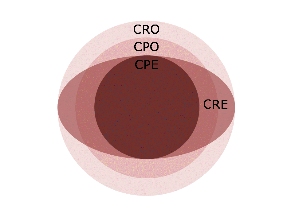
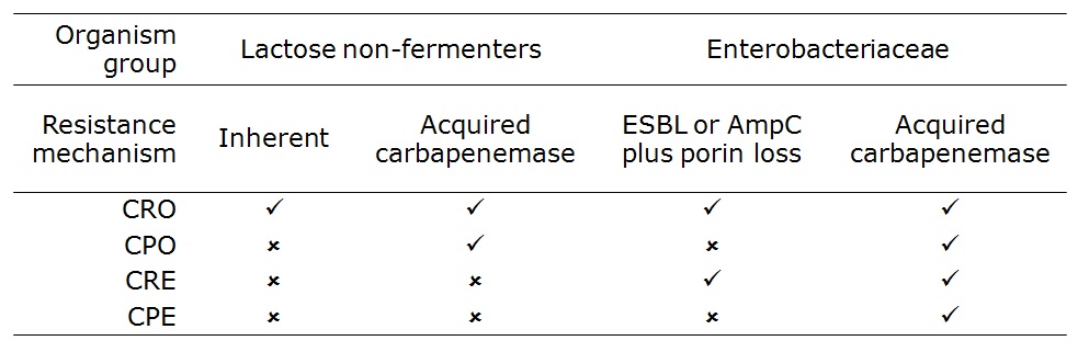
Control of hyperendemic Carbapenem-resistant Enterobacteriacae

Abdul Ghafur – The Indian perspective

A passionate presentation by Dr Ghafur beginning with 50% attributable mortality in his neutropaenic patients due to CRE resulting from carbapenem and colistin abuse; mortality for pan resistant CRE approached 100%. Dr Ghafur discussed some of the underlying problems in India: poor sanitation, high rates of carriage of resistant bacteria (80% CR Acinetobacter and 40% CR Klebsiella), reflecting on the fact that tight local antimicrobial prescription will not save you if everyone else is sloppy. How to treat pan-drug resistant CRE? “Pentatherapy” using a 5 drug cocktail and a prayer. (Actually, there is probably some synergy between colistin and other antibiotics so pentatherapy isn’t as daft as it sounds.)

Yehuda Carmeli An Israeli perspective

Israel experienced a dramatic initially clonal national outbreak of CRE (KPC CC258) in 2006. Analysis of data from the outbreak indicated that compliance with cohorting correlated tightly with CRE transmission; more data that getting the basics right works. The risk-based screening that was implemented detected a low prevalence of carriage (5% from long-term care facilities, 0.5% from other high risk groups) but was considered worthwhile to prevent outbreaks. Dr Carmelli raised several points for discussion:

  • Media interest. It seems to me that the media can be a friend or foe in dealing with outbreaks and wider healthcare issues. If left unbriefed, they will be a foe. But if properly briefed, they can be a powerful ally.
  • Emergence of different CRE genes (OXA-48 and NDM-1) in Israel with different epidemiological associations than KPC.
  • Dealing with the long-term care facility reservoir.
  • Non-compliance with basic infection control.
  • “Eye off the ball” syndrome, where hospital administrators only respond to current threats.

The national successful response in Israel is an encouraging success story. However, Israel has a population of 8 million, with only 31 hospitals. Can successful national control strategies be implemented in larger countries with an inferior healthcare infrastructure and national debt problems such as Greece and Italy?

Achilles Gikas – The Greek perspective on CRE: from surveillance to control

Dr Gikas described the development of the CRE epidemic in Greece with overlapping outbreaks of VIM and KPC carriers, and how CRE is now a “frequent flyer” travelling around the world. Reassuringly, there are some local success stories in Greece where focused interventions have reduced rates of CRE, but, mindful of Dr Kulakkattil’s comment that “no hospital is an island”, concerted national efforts are required to grasp this rampaging Hellenic bull by the horns.

Environmental decontamination with hydrogen peroxide vapor – does the effect evaporate over the Atlantic? Perl v Huttner

Having spent some 10 years researching this particular topic, needless to say I was looking forward to this debate. The debate was not particularly well framed. Was this about most of the studies of HPV (and all of the controlled studies with a clinical outcome) coming from US hospitals? Or that many European countries have a lower proportion of single rooms, which makes the application of HPV more challenging? Or a wider debate about whether to consider “no-touch” disinfection (NTD) systems at all? I felt that Dr Perl addressed the more general questions while Dr Huttner focused on HPV studies, which rather took the wind out of the sails of the debate.

Dr Perl summarized data that pathogens are shed into the environment, they survive for extended periods, persist despite conventional cleaning and disinfection and improving conventional methods helps, but transmission continues. Hence, NTD systems and specifically HPV are warranted in some circumstance. Dr Perl acknowledged the lack of RCTs but showed convincing data that HPV eliminates pathogens from surfaces and reduces transmission, and fulfils Bradford-Hill’s criteria of causation.

Dr Huttner began like Dr Perl by considering some things that evaporate over the Atlantic: big cars, big guns and flavoured coffee to name but a few! His American wife gives him an unusually sharp perspective on trans-Atlantic issues. Dr Huttner had clearly done his homework and read the various HPV studies in detail. He presented a series of good points, although I didn’t feel that he constructed a particularly coherent argument. To address some specific points:

The short debate (30 mins) did not allow time for the authors to present a rebuttal, which was a shame, but it was a stimulating session.

FRI 28th JUNE

Controlling ESBLs – a global perspective

Jean-Christophe Lucet – Europe

Dr Lucet began by demonstrating a startling increase in the proportion of invasive isolates that carry ESBLs in Europe comparing 2005 with 2011. He reviewed data that ESBL-producing K. pneumoniae is much more likely to be transmitted in hospitals than ESBL-producing E. coli, perhaps due to an increased capacity for environmental survival. Effective interventions are difficult to recommend due to lack of RCTs, outbreak settings (and regression to the mean), publication bias and control bundles. However, Dr Lucet concluded:

  • Hand hygiene: yes.
  • Active surveillance cultures and contact precautions: equivocal.
  • Cohorting: yes during outbreaks.
  • Environmental control: less important than for other pathogens.
  • Selective digestive decontamination (SDD) / universal use of chlorhexidine: equivocal. My view is that SDD should not be used – the indiscriminate use of antibiotics will drive further resistance.

Wing-Hong Seto – Far East / Asia

Carriage rates of ESBLs in E. coli and K. pneumoniae are alarmingly high in parts of Asia, reaching >50% in India and Vietnam. Dr Seto suggested that single room isolation is more important for Gram-positive vs. Gram-negative bacterial pathogens based on relatively higher recovery of Gram-positive bacteria from the hospital environment. However, lactose non-fermenting Gram-negatives such as Acinetobacter seem to ‘mimic’ Gram-positive bacteria in terms of their environmental survival, and even the Enterobacteriaceae can survive for days to weeks to months on surfaces. So, I would argue that priority for single room isolation should be dictated by an assessment of local epidemiology and risk, not the survival properties of various bacteria.

Andrew Stewardson – Australia

Carriage rates of ESBL-producing bacteria are lower in Australia than in nearby Asian countries (<10% in the hospital population and <5% in the community). Dr Stewardson presented some interesting Australian data showing that rigorous implementation of standard precautions and antibiotic restriction programmes can result in impressive reductions of ESBL producing K. pneumoniae in hospitals.

Final reflections

My first ICPIC was an enjoyable experience. Highlights included the opening session learning lessons from SARS, the entertaining innovation academy and some grim updates on the ‘rampaging Hellenic bull’ that is CRE. Regretfully, I didn’t get to see a single poster and missed few concurrent sessions (including the review of best papers by Eli Perencevich and Andreas Widmer – they’ve published their slides here). Of all the analogies presented at the conference, I think the most useful was the ‘Infection Control Football Club’ by Dr Sheldon Stone. Hand hygiene is the goal keeper (i.e. last line of defence). But you won’t have a winning football team with just a goal keeper. No matter how good they are, they will never stop every shot.