How much patient-to-patient spread of S. aureus occurs? Apparently, not much

I recently posted an article on surprising finds of a study suggesting that horizontal transmission of C. difficile from known symptomatic cases may be less common that we thought. A group of researchers from Oxford, Brighton and London in the UK applied similar methodology to Staphylococcus aureus transmission with similar findings: only a fifth of S. aureus acquisitions could be attributed to patient-to-patient transmission.

All patients admitted to a 16 bed ICU in Brighton were screened on admission and weekly to detect S. aureus colonization and acquisition. Each isolate was typed by spa and whole genome sequencing (WGS). The number of acquisitions that could be linked using conventional methods (spa typing combined with an analysis of overlapping stays) vs. WGS was evaluated.

Overall, 185 (16.7%) of 1109 admissions carried S. aureus; 59 carried MRSA (5.3%). 680 patients were on the unit for long enough to have a weekly screen and hence were eligible for assessing acquisition. Of these, 44 S. aureus (22 MRSA) acquisitions were detected in 41 patients. 35 of these acquisitions were in patients who were screen-negative on admission and 9 were acquisitions of different strains in patients who were already colonized on admission.

Only 14% (5/36) of the acquisitions available for typing were of the same spa type as another patient on the unit at the same time. All of these were MRSA. WGS discounted three of these apparent occurrences of patient-to-patient transmission, confirmed two and identified a further five (3 MRSA, 2 MSSA). So, in total, 7 / 36 (19%) of acquired isolates (5 MRSA and 2 MSSA) were linked to isolates from other patients (Figure).

Price MRSA WGS
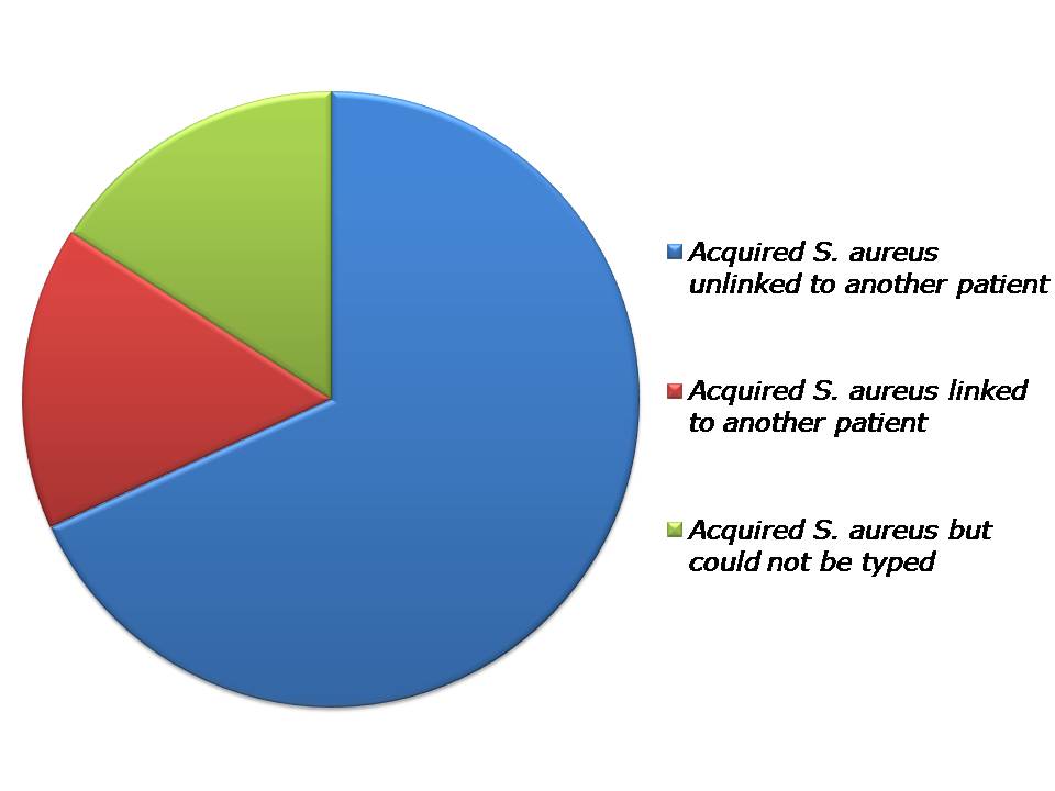
Figure: Source of S. aureus acquisitions identified through WGS.

The paper raises some interesting questions:

  • Principally, if almost 80% of patients did not acquire their S. aureus from other patients on the unit, where on earth did they acquire them from? In the C. difficile study, unrecognized importation of diverse strains from the community that would not be detected on admission since admission screening was not performed represents a plausible explanation for the surprisingly low incidence of horizontal transmission from known cases. This is not the case in this study, where all patients were screened on admission for S. aureus. So where were the diverse isolates acquired from? Staff carriers? Contaminated surfaces? S. aureus has the capacity to survive on surfaces for very long periods (more than 1 year), so an ancient environmental reservoir is possible. Furthermore, there was no ‘lead in’ period to the study, so it could be that S. aureus on the unit in the months before the study left an environmental reservoir that led to acquisition in some cases, which would not have been captured by this study. The fact that 4/7 patient-to-patient transmissions acquired the same strain of S. aureus without sharing time on the ICU together supports this, although could also be explained by staff carriers. It was a shame that 16% of the 329 S. aureus isolates (including 16% (7/44) of the acquired isolates) were not available for sequencing, which represents a substantial reservoir from which patient-to-patient transmission likely occurred in some cases.
  • It was interesting that some 20% (9/44) acquisitions that were detected occurred in patients who were already colonized on admission; these would have been missed altogether if molecular typing was not performed. I wonder how much ‘silent’ acquisition of this type occurs?
  • Assuming temporal relationship between strains assumes a constant mutation rate. The ‘speed of the mutation clock’ was assessed in this study through repeated sampling of the same patient. This exercise demonstrated minimal diversity, as was the case for C. difficile.
  • WGS is rapidly becoming the gold standard for transmission mapping. In this study, the conventional approach of evaluating spa typing with overlapping stays lacked both sensitivity and specificity for identifying transmitted isolates.

In summary, the major finding is that only 20% of patients who acquired S. aureus appeared to acquire it through horizontal spread from other patients. The next frontier of transmission mapping must be a more comprehensive evaluation of other potential sources: contaminated surfaces, contaminated air, nurses, doctors, cleaners, tea-tray deliverers and the list goes on…

Article citation: Price JR, Golubchik T, Cole K, Wilson DJ, Crook DW, Thwaites GE, Bowden R, Walker AS, Peto TE, Paul J, Llewelyn MJ. Whole-genome sequencing shows that patient-to-patient transmission rarely accounts for acquisition of Staphylococcus aureus in an intensive care unit. Clin Infect Dis 2014. Jan 9. [Epub ahead of print].

How much Clostridium difficile infection is hospital-acquired?

B0006630 Clostridium difficile

This is a very impressive New England Journal of Medicine study from an Oxford University based group, using whole genome sequencing to really dissect relatedness of C. difficile isolates over a 5 year period. The study evaluates how many cases of C. difficile infection (CDI) were caused by isolates that were genetically related to previous symptomatic cases. This is not quite the same thing as evaluating how much CDI is hospital-acquired, mainly because the test used to detect CDI in the study has been phased out due to poor sensitivity, patients and staff were not screened for asymptomatic C. difficile carriage, and the environment was not sampled, so there was a large, unrecognized, hospital-based C. difficile reservoir from which horizontal transmission almost certainly occurred.

A major problem was the use of an Enzyme Immuno Assay (EIA) test kit to detect CDI. Whist these tests were used pretty much universally in the UK at the time of the study, they have now been shown to be very unsatisfactory. The sensitivity of EIA for the detection of CDI has been as low as 50% in some studies. Put another way, for every case of CDI that is detected, one goes undetected. This is crucially important in the context of this study, where the undetected CDI cases would contribute to the burden of asymptomatic carriers, which together would contribute to transmission. It’s also worth noting that C. difficile could not be cultured from 25% of stool samples that were EIA-positive, suggesting that the test may have had poor specificity too. The authors did try to ‘control’ for this problem, by effectively assuming that all stool specimens tested for CDI were positive in a sensitivity analysis, but this did not really help in explaining genetically related cases with no discernable epidemiological links.

There is also a technical point about the definition of ‘genetically distinct’ in terms of whole genome sequencing. If two isolates differ by 11 base pairs across the whole genome, do they originate from the same strain? It’s difficult to tell. In this study, they used a fairly conservative measure of relatedness: >10 single nucleotide variants (SNVs) was considered ‘genetically distinct’, and ≤2 SNVs was considered ‘genetically related’. This may have over-estimated apparent genetic heterogeneity. To be fair, the authors did perform a careful ‘validation’ study to determine the clock speed of mutation in their isolates by sequencing the first and list isolates obtained from a sample of patients, finding that 0-2 SNVs were expected for isolates <124 days apart. Even using these conservative measures of relatedness, the majority (55%) of isolates were related (‘not genetically distinct’ to be precise) to others in the collection (≤10 SNVs) and around a third of isolates were ‘genetically related’ to others in the collection (≤2 SNVs).

The authors performed detailed work to explore epidemiological associations between genetically related isolates (Figure). No acute- or community-based epidemiological links could be identified for 36% of the 333 genetically related cases, which perhaps supports the presence of unrecognized symptomatic cases or asymptomatic carriers.

CDI eyreFigure: Epidemiology relationships between 333 genetically related cases. ‘Ward contact’ = shared time on the same ward; ‘Hospital contact’ = shared time in the same hospital, without direct ward contact; ‘Ward contamination’ = admitted to the same ward within 28 days of the discharge of a symptomatic patient; ‘Same GP’ = no hospital contact, but shared the same GP; ‘Same postcode’ = no hospital contact, but shared the same postal code).

The overall rate of CDI was low, at <1 per 1000 patient days and it is noteworthy that the prevalence of genetically related and genetically distinct cases declined during the study period. I suspect if the same study had been performed for the period of 2000-2005, when more hospital transmission was almost certainly occurring, then a far higher proportion of isolates would have been genetically related.

I fear that this study will be used by some to ‘prove’ that horizontal transmission of C. difficile in healthcare settings is now uncommon, and most hospital-onset cases can be explained away by “CA-CDI”. Due to the poor sensitivity of the diagnostic kit combined with the likelihood of asymptomatic human carriage and environmental contamination, this study does not answer the question of how much CDI is hospital-acquired. It does, however, suggest that horizontal transmission from known symptomatic cases may be less common that we thought.

Article citation: Eyre DW, Cule ML, Wilson DJ et al. Diverse sources of C. difficile infection identified on whole-genome sequencing. N Engl J Med 2013; 369: 1195-1205.

Photo credit: Annie Cavanagh. Wellcome Images.

Is there a causal relationship between contamination burden and transmission risk?

contamination v transmission There’s an age-old problem in science: how do you prove a causal relationship between variables that correlate? Proving that the variables are correlated is the easy part; it’s more difficult to disentangle cause from effect. This can be seen in several studies that identify a correlation between environmental burden and the number of patients that are infected or colonized with pathogens.DentonFigure 1. Correlation between the number of patients infected with Acinetobacter spp. and the number of positive Acinetobacter spp. environmental cultures per calendar month during an outbreak on a neurosurgical ICU.1

SalgadoFigure 2. Correlation between microbial burden and the number of patients who acquired an HAI in ICUs.2

WhiteFigure 3. Correlation between the number of hygiene failures and the number of patients who acquired an infection on a surgical intensive care unit each week.3

So can we conclude that the higher burden of contamination resulted in an increased risk of acquisition? Or is it that more patients were infected or colonized with pathogens, which resulted in more environmental shedding? From these studies, you can’t be sure.

If you were seeking to prove the role of a gene in a process, you’d knock out the gene and demonstrate that the process stopped or changed. So, the only way to disentangle cause and effect in contamination and transmission is to perform an intervention to reduce environmental contamination and show that this correlates with reduced transmission. While the Salgado study evaluated an intervention, the data correlating contamination burden with HAIs was not stratified by the intervention, which would have been one way to assess likely causation.2

There is some further in vitro and epidemiological data supporting that the degree of transmission may be proportional to the environmental burden. An in vitro mouse model established a ‘dose-response’ relationship between the degree of contamination with C. difficile spores and the development of CDI.4 Furthermore, this model showed that disinfectants that achieved a greater log reduction of C. difficile spores were more able to interrupt transmission.

Also, one of the studies demonstrating that admission to a room previously occupied by a patient with VRE increases the chances of VRE acquisition identified something amounting to a ‘dose response’.5 The greatest increased risk was for patients admitted to a room with an environmental culture positive for VRE, and being admitted to a room where the immediate prior room occupant was colonized with VRE carried a greater increased risk than being admitted to a room where any patient in the 2 weeks prior to admission was VRE colonized (Figure 4).

DreesFigure 4. How the increased risk of acquiring VRE from the prior room occupant changes due to patient and environmental factors.5

Is there a causal relationship between contamination burden and transmission risk? On balance, the answer seems to be yes, though it would be useful to have a solid intervention study to prove that an increasing environmental burden causes an incrementally increase in transmission risk.

Article citations:

  1. Denton M, Wilcox MH, Parnell P et al. Role of environmental cleaning in controlling an outbreak of Acinetobacter baumannii on a neurosurgical intensive care unit. J Hosp Infect 2004; 56: 106-110.
  2. Salgado CD, Sepkowitz KA, John JF et al. Copper surfaces reduce the rate of healthcare-acquired infections in the intensive care unit. Infect Control Hosp Epidemiol 2013; 34: 479-486.
  3. White LF, Dancer SJ, Robertson C, McDonald J. Are hygiene standards useful in assessing infection risk? Am J Infect Control 2008; 36: 381-384.
  4. Lawley TD, Clare S, Deakin LJ et al. Use of purified Clostridium difficile spores to facilitate evaluation of health care disinfection regimens. Appl Environ Microbiol 2010; 76: 6895-6900.
  5. Drees M, Snydman D, Schmid C et al. Prior environmental contamination increases the risk of acquisition of vancomycin-resistant enterococci. Clin Infect Dis 2008; 46: 678-685.

The terms 'horizontal' and 'vertical' intervention leave me feeling upside down, confused

horizontal vertical

I am no expert in HIV, but I know that ‘vertical transmission’ means something very specific:

Vertical transmission: the transmission of a disease from mother to child either during pregnancy, childbirth, or by breastfeeding.

Similarly, the definition of ‘horizontal transmission’ is well defined:

Horizontal transmission: the transfer of an infection from person to person.

So, when I read about ‘vertical’ and ‘horizontal’ interventions in a recent New England Journal of Medicine Editorial and the Controversies blog, I began to get a little confused. I have a PhD in epidemiology so don’t consider myself easy to confuse (in this particular domain), but I would have thought that a ‘horizontal intervention’ would be directed towards preventing horizontal spread of an infectious agent and a ‘vertical intervention’ would be directed towards preventing the vertical transmission of an infectious agent. But this is not how these terms are being applied. Instead, a ‘horizontal intervention’ is being used to describe an intervention applied to every patient (such as chlorhexidine bathing or hospital-wide hand hygiene interventions) whereas a ‘vertical intervention’ is being used to describe an intervention designed to reduce colonization or infection due to a specific pathogen (such as active screening and isolation to prevent the spread of MRSA). The use of the term ‘vertical intervention’ seems especially confusing, since it’s a ‘vertical intervention’ to prevent the horizontal transmission of a specific pathogen!

I fail to see how the terms ‘vertical’ or ‘horizontal’ intervention are useful when there are such well-established definitions for horizontal and vertical transmission. I think that ‘universal intervention’ (such as universal screening or decolonization) and ‘targeted intervention’ (such as active screening and isolation to prevent the spread of MRSA) make a lot more sense. These terms are already in common circulation, so I would urge those who favour the use of ‘vertical’ or ‘horizontal’ intervention to reconsider their terminology.

MDR-Acinetobacter baumannii beats MRSA in the war for ICU predominance

A. baumannii is a notorious nosocomial pathogen due to a combination of its environmental resilience, its association with antimicrobial resistance and its outbreak potential. Colonized patients and contaminated environments are thought to be the primary reservoirs for the nosocomial transmission of this pathogen.

A recent study from China suggests that carriers of MDR A. baumannii (MDR-AB) show stronger ability to contaminate their immediate environment than those carrying MRSA and that MDR-AB spreads more easily and rapidly among inpatients compared with MRSA. The 20-month study was conducted in a respiratory ICU (RICU) where active screening of patients and targeted environmental screening for MRSA and MDR-AB were performed. The environmental samples were collected from 6 sites on patients’ bed linens.

High levels of carriage and nosocomial acquisition were found among the 175 patients admitted to the RICU where 44% of the patients were MDR-AB positive (80% of which were hospital acquired) and 24% of patients were MRSA carriers (60% of which were hospital acquired). Interestingly, 15.4% of the patients were co-carriers of MRSA and MDR-AB.

Researchers found that bed linens were commonly contaminated with MRSA and MDR-AB and that the contamination rate for MDR-AB was significantly higher than that of MRSA. Of the 576 MRSA samples, 26.6% were positive, and 51.6% of the 1,176 MDR-AB swabs were positive. This is surprising given the strict daily extensive cleaning practices, thrice daily bed linen changes and stringent terminal sterilization immediately after discharge of carriers. Researchers used the weekly colonisation pressure adjusted by degree of bed linen contamination (WCPe) and weekly acquisition rate (WAR) as parameters to evaluate the potential spread of these pathogens among inpatients. They found a positive significant correlation between the WCPe and WAR values for both organisms but both the WCPe and WAR of MDR-AB were significantly higher than for MRSA.

This study shows that environmental contamination with MDR-AB and the rate of its nosocomial acquisition is significantly higher than those for MRSA, which may explain why MDR-AB is able to spread among inpatients more rapidly. Although the study found positive significant correlation between the WCPe and WAR in the subsequent weeks, this correlation does not necessarily indicate causality. Nevertheless, the authors conclude that reduction of environmental contamination close to MDR-AB positive patients is crucial in controlling MDR-AB transmission.

Article citation:

Sui W, Wang J, Wang H et al. Comparing the transmission potential of Methicillin-resistantStaphylococcus aureus and multidrug-resistant Acinetobacter baumannii among inpatients using target environmental monitoring. Am J Infect Control. 2012. doi: 10.1016/j.ajic.2012.08.007