Are beards bug traps? Au contraire!

As the owner of a relatively new beard (see picture below), I was alarmed to hear that my beard is probably as contaminated with faeces as a toilet brush. Fortunately, a Journal of Hospital Infection study from 2014 turns this on its head, showing that those wearing beards are actually less likely to be colonised with staphylococci!

Me and my beard

Twitter01f6551v2

Continue reading

The inanimate environment doesn’t contribute to pathogen transmission in the operating room…OR does it?

OR

Nosocomial or hospital-acquired infections are a worldwide problem affecting millions of patients yearly and increasing morbidity and mortality. The role of the hospital inanimate environment (environmental surfaces and surfaces of medical equipment) in the transmission of certain nosocomial pathogens such as C. difficile, norovirus, MRSA, VRE and Acinetobacter is now well established supported by various studies and publications. Most, if not all of these studies, investigated the transmission process in patient rooms or ICUs. Although the role of air in the transmission of pathogens has been extensively studies in the operating room (OR) setting, do contaminated surfaces play a role in pathogen transmission in the OR?

A recent review article published in the journal “Surgical Infection” questioned whether the OR inanimate environment contributed to the transmission of pathogens, hence possibly causing infections including surgical site infections (SSIs). Few studies have investigated surface contamination in the OR and even fewer have investigated possible pathogen transmission from the environment in this setting. While the inanimate environment in the OR has been considered a potential source for pathogens that may cause SSIs for more than 100 years, the role of this environment in the patient acquisition process within this setting is still debatable. Before revealing the conclusions of the review paper, I would like to look at both sides of the argument.

THE OR INANIMATE ENVIRONMENT DOES NOT PLAY A ROLE IN PATHOGEN TRANSMISSION AND INFECTION

The patient population and length of stay

In a hospital, patients colonised or infected may spend days or even months in ward rooms or ICUs increasing the chance that these patients will contaminate their environment or acquire pathogens from that environment. The likelihood of environmental contamination or pathogen acquisition increases with the length of hospital stay as well as other factors such as gross contamination and soiling.

In the OR setting however most patients spend only few hours under full or partial anaesthesia. This makes it less likely that these patients will contaminate their environment or acquire pathogens from the environment (by self inoculation at least). In addition, although gross contamination via blood for example is common, other type of gross environmental contamination linked to transmission such as diarrhoea and vomiting are less likely to occur in an OR.

The OR environment (surfaces and air)

Unlike most patient rooms, OR air quality is well regulated to prevent contamination via the air. This not only reduces the risk of infection via airborne pathogens but also reduces the amount of pathogens settling on and contaminating environmental surfaces in ORs. In addition, the OR inanimate environment is routinely cleaned/disinfected. Most ORs are cleaned at the end of the working day and many surfaces and areas are cleaned before and between surgeries with strict policies on how to deal with gross contamination (e.g. blood and tissue).

Minimising infection risk

As most SSIs are thought to originate from patients’ or healthcare personnel’s own flora, many interventions are in place in ORs to minimise the risk of contamination and infection. These include policies for hand scrubbing and disinfection, gloving, masks, and the proper preparation of patients’ skin before incision. The instruments used in surgery are also routinely sterilised before surgery to minimise the risk of infection.

Organisms involved in SSIs

The hospital environment has been implicated in the transmission of a number of pathogens including norovirus, C. difficile, MRSA, VRE and Acinetobacter. These pathogens are able to contaminate the environment at a high load and survive for long period of time facilitating transmission and acquisition.  While infections with these organisms can be acquired in the OR, with the exception of Staphylococcus species, these pathogens are not the major causes of SSIs. The environmental resilience of other organisms involved in SSIs is not well characterised and it is unclear whether they can survive long enough in the environment to be transmitted.

THE OR INANIMATE ENVIRONMENT IS A SOURCE OF PATHOGENS THAT CAUSE INFECTION

The OR environment

ORs are busy, with many personnel involved during a surgical procedure, some of whom come and go in and out of the OR during the process. It is also an environment with multiple and frequent contact between personnel, patients and the environment including medical equipment. It is difficult if not impossible to observe the WHO’s 5 moments for hand hygiene in such an environment, or to clean and disinfect the environmental surfaces effectively during a surgical procedure. Organisms originating from the floor of the OR can also be disturbed by walking and are taken into the air which may increase the risk of infection.

The OR inanimate environment is contaminated

Many people in the general public think of ORs as ultra clean, even sterile, environments. For anyone working in ORs, it is clear that this view is far from the truth. Although modern ORs have strict measures to reduce contamination, the OR inanimate environment becomes contaminated with various organisms including those involved in SSIs. Studies have reported contamination of various OR areas such as anaesthesia equipment, beds, intravenous pumps and poles, computer keyboards, telephones and OR floors.  A variety of pathogens capable of causing infections have been identified including Gram-negative bacilli such as Acinetobacter and Pseudomonas species, Staphylococcus including (MRSA) and Enterococcus. These results may be in part due to the fact that suboptimal cleaning in ORs is a widespread issue in hospitals.

Pathogen transmission occurs in ORs

A number of studies in ORs focusing on the role of anaesthesia equipment and providers in the contamination and transmission of pathogens in ORs have concluded that the hands of anaesthesia providers, patient IV tubing and the immediate patient environment were contaminated immediately before or during patient care with a wide range of bacterial pathogens leading to transmission. Transmission of pathogens from and to the hands of the anaesthesia providers involving the inanimate environment occurs frequently given the frequent contact with the environment in ORs.

Human behaviour in ORs contributes to environmental contamination and transmission

We are all familiar with the view that surgeons tend to be the worst healthcare workers as far as hand hygiene compliance is concerned. However, this is only the tip of the iceberg regarding lapses in infection prevention in ORs. For instance, anaesthesia provider’s behaviour and attitude including confusion on when and how often to perform hand hygiene during a procedure is a common cause of pathogen transmission. In one study, anaesthesia providers touched 1,132 objects during 8 hours of observations in OR, but only performed a total of 13 hand disinfections. No hand disinfections were witnessed at any time during 3 (43%) of the procedures observed. Furthermore, hand hygiene failed to precede or follow procedures, blood exposure or contact with the floor. Alarmingly, it has been reported that objects that fall onto the OR floors during surgery were frequently placed back either on to horizontal work surfaces or even on to the patients themselves during operations.

THE CONCLUSION

It is clear that the inanimate environment of the OR, including medical equipment, can become contaminated with pathogens that cause infections including SSIs. These pathogens can then be transmitted to the hands of healthcare workers and have the potential to cause infection. Further studies are necessary to quantify the role of contaminated surfaces in the transmission of pathogens and to inform the most effective environmental interventions in the ORs. Given the serious consequences of SSIs, special attention should be given to the proper cleaning and disinfection of the inanimate environment in ORs in addition to the other established measures to reduce the burden of SSIs. These include addressing the human behaviour that contributes to environmental contamination and transport of surface pathogens into the vulnerable sites of patients during surgery. Such measures include reducing human traffic in ORs, stricter adherence to the standard operating protocols during procedures, and compliance with proper hand hygiene and gloving. Specific hand hygiene guidelines tailored to OR personnel may be needed given the large number of hand contact events per hour in these settings.

Image: NIH Library.

Reflections from HIS 2014, Part II: Dealing with the contaminated environment

HIS_Web_Banner_Jpeg

Welcome to Part II of my reflections from HIS. For the box-set, see the list at the beginning of Part I here.

Dr Karen Vickery – Multispecies biofilms on dry hospital surfaces – harbouring and protecting multiantibiotic resistant organisms

Probably the most important update from the entire conference was more data from the Vickery lab on biofilms on dry hospital surfaces. She excised 44 dry surface samples from the ICU, put them under the electron microscope and, lo and behold, 41 of them (93%) had fully-fledged (if somewhat unusual) EPS-producing biofilms on! The implications are huge: this could explain extended surface survival, poor success rate of surface sampling, and result in reduced biocide susceptibility up to the tune of 1000x (see my review just published in JHI with Karen as a co-author for more on biocides and biofilm susceptibility).

Dr Silvia Munoz-Price – Controlling multidrug resistant Gram-negative bacilli in your hospital: We can do it so can you!

Dr Munoz-Price described her hospital’s impressive reductions on carbapenem-resistant A. baumannii – from 12 new isolates per week to virtually none today. So what worked? It’s difficult to be sure since it was a bundled intervention. Dr Munoz-Price described the rationale behind some elements of the bundle: environmental surface and staff hand sampling to visualize the invisible, environmental cleaning and disinfection to deal with the ‘fecal [sic] patina’ [a stooly veneer emanating from the rectum] (see Dr Munoz-Price and Dr Rosa’s guest blog for more details), and chlorhexidine bathing. Perhaps the most interesting aspect was the various implementation challenges that were overcome. It was amazing how far removed practice ‘in the trenches’ was from the policy set by the epidemiologist’s office, exemplified by environmental staff buying their own UV lamps to for “spot cleaning” removal of fluorescent markers of cleaning thoroughness. Overcoming these challenges required more that the stick (citations for non-compliance, which failed); culture change takes understanding, time and a very large carrot (and some sticks too, sometimes).

Jim Gauthier – faeces management

A number of key pathogens are associated with faecal colonization and shedding: C. difficile, VRE, ESBL and CRE. Jim didn’t mention MRSA, but this can also cause gastrointestinal colonization and, more controversially, infection. Enterobacteriaceae can survive on dry surfaces for longer than you’d expect, too. We traditionally worry about surface contamination of high-touch sites in inpatient settings. Floor contamination isn’t important (unless you happen to be a wheel chair user, a toddler, or drop your pen). Contamination in outpatient settings isn’t a problem either (unless you happen to have a fairly short consultation for a patient with VRE). So, what to do? Jim introduced the idea of a ‘hierarchy of control’; put another way, prevention is better than cure, so do we have the right systems in place to manage faeces which is teeming with hospital pathogens? For example, should we be enforcing mandatory contact precautions for all contact with faeces (standard precautions – which aren’t very standard anyway – are probably not adequate)? Finally, Jim mentioned the growing importance of faecal microbiota transplantation (and hearing a Canadian speak about this reminded me of a hilarious spoof video).

No-touch automated room decontamination (NTD)

medical equipment in a hospital roomFigure: Hospital bed rails are frequently contaminated, and often not easy to clean and disinfect using conventional methods. 

Paul Dickens – establishing Ebola surge isolation capacity in the UK

Paul Dickens gave a whistle-stop overview of the detailed plans for Ebola surge capacity in the UK (perish the thought). He began by describing the replacement of formaldehyde with hydrogen peroxide vapour for the decontamination of the patient isolators at the Royal Free High Level Isolation Unit (HLIU). They now have a tried and tested process and protocols in place to get the HLIU back online within days using hydrogen peroxide vapour decontamination, where the previous protocol using formaldehyde put it out of action for 6 weeks! (I was involved in writing the protocols for this tricky decontamination assignment, which were reported on a poster published at HIS.) Other challenges in establishing surge capacity include staff expertise, and PPE recommendations, supply & training. Surge capacity is now established. Let’s just hope we won’t need it!

Dr Frédéric Barbut – How to eradicate Clostridium difficile spores from the environment

There’s now plenty of evidence that contaminated surfaces contribute to the transmission of C. difficile. These environmental intervention studies show a 50-80% reduction in the rate of CDI; does this mean that 50-80% of CDI acquisition is environmentally-associated? This seems too high, but it’s difficult to think of another explanation. Furthermore, there is emerging but compelling evidence of a proportional relationship between the degree of C. difficile surface contamination and transmission risk? I really don’t think that the public have yet ‘got’ that the previous occupant can influence acquisition risk. And when they do, I think there will be increasing demand for properly decontamination rooms. So, is it time to turn to NTD systems? Sometimes, yes. And do you go for hydrogen peroxide or UV? Well, that depends on what you’re trying to achieve! If you’re trying to eliminate pathogens, which sometimes you will be, then hydrogen peroxide vapour is the best choice. But if you’re trying to reduce contamination levels without necessarily eliminating all pathogens, then UV is the best choice due to its speed and ease of use.

The debate: “Hospitals that do not use high-tech decontamination of the environment are doing their patients a disservice.”

This debate pitted Profs Hilary Humphreys and Phil Carling (pro) against Peter Hoffman and Martin Kiernan (con). It was lively, entertaining and engaging…

Prof Humphreys argued that it is not acceptable to admit patients to rooms with inherent additional risk for transmission. We can address this by ‘walking like the Egyptians’ and copperising our surfaces, for which there is now some data with a clinical outcome. Another approach is NTD systems, for which data (including some clinical outcomes) are emerging. Prof Carling’s presentation was somewhat unusual, with his arguments seemingly an appeal to common sense rather than drawn from the published literature.

Martin Kiernan began by acknowledging the role of the environment, but that hand contamination is almost always the final vector (and there’s some evidence for this). The cornerstone of Martin’s argument was that whether NTD systems work is the wrong question. We should be focusing our time, money and attention on improving conventional methods which have been shown to reduce transmission. Peter Hoffman complemented Martin’s pragmatic viewpoint with thorough, thoughtful critiques of the studies on HPV decontamination with a clinical outcome. The 2008 Boyce study has more holes than the 2013 Passaretti study, which itself is far from watertight!

The key argument for turning to NTD systems is that admission to a room previously occupied by a patient with an MDRO increases the risk of acquisition due to residual contamination, and NTD decontamination mitigates this increased risk. So, my own conclusion is that hospitals that do not use high-tech decontamination of the environment are indeed doing their patients a disservice. Sometimes!

Look out for the third and final installment of my reflections from HIS 2014 at some point tomorrow!

Image: Medical equipment in a hospital room.

HIS Poster Round: Dealing with contaminated hands, surfaces, water and medical devices

poster round

I was delighted to be asked to lead a poster round at the Healthcare Infection Society (HIS) conference. I was a bit disappointed to see that the poster sessions are tacked on to the end of the day (when everybody’s had enough and really just wants to retreat to their hotel room for an hour before the evening’s activities). My view is that posters are the lifeblood of conferences (and I am not alone in this view); they should have much more prominence, with a “ringfenced” session integrated into the main program.

Anyway, whinge over, I thought I’d share which posters I chose, why I chose them, and what I want to know about them.

You can access the abstracts here.

GENERAL CATEGORY

#3200 Longhurst et al. Hand drying methods in NHS England Trusts, September 2013.

I chose this poster because it’s becoming increasingly clear that the choice of hand drying method can influence the degree of bacterial contamination. Also, whilst I accept the economic and environmental benefits of jet and warm air dryers, they always seem to leave my hands a bit damp. (Perhaps I just have sweaty palms.) Anyway, this is what I want to know about this poster:

  • Why did you feel the need to ‘enforce’ a response by using the Freedom of Information act?
  • Why do you think jet / warm air dryers were rare in clinical areas?
  • Do you think that jet and to a lesser extent warm air dryers result in dangerous dispersal of microbes?

#3349 Tang et al. A 3 year hand hygiene program to increase compliance rate for heatlhcare providers in the A&E Department of Tuen Mun Hospital in Hong Kong.

There’s not a lot of data on hand hygiene compliance in A&E. A 2005 study examined compliance with hand-washing in the TV show ER, reporting a hand hygiene compliance rate of 0.2%. Yep, that’s ZERO POINT TWO PERCENT! Although reality is marginally better (according to this review), there’s work to be done, so I chose this poster mainly because of the impressive impact in improving hand hygiene compliance. My questions are:

  • Which of the barriers to hand hygiene that you mention do you think is most important?
  • Who was on your task-force to decide what to do?
  • How many people completed the questionnaires?
  • How many observations were done in each time period?
  • How do you measure that the awareness of hand hygiene increased?

#3174 Khanafer et al. Hospital management of Clostridium difficile infection: a literature synthesis

This is a novel review of the literature: using the ORION checklist to capture variables that help us to determine what works to control CDI from outbreak reports and intervention studies. Here’s what I want to know:

  • Can we really derive anything useful about which intervention works when you have more than one variable, even if studies are reported in a structured way? (High school science is pretty clear: change one variable at a time!)
  • How can these % reductions be so high when (apparently) only 30% of CDI is hospital-acquired (according to some people’s interpretation of the Oxford WGS difficile study)?
  • Which is the single most important intervention to prevent CDI transmission?

ENVIRONMENT CATEGORY

#3285 Cunningham et al. VRE Outbreak Control – the Need for Speed (Use of Molecular Technology)

Two of my favourite subjects: VRE and rapid diagnostics! Here are my questions:

  • Why bother trying to control VRE? Some pretty persuasive voices are arging that it’s not worth it!
  • Are you sure that rapid diagnostics made the difference? You also introduced enhanced cleaning / disinfection, extra screening, pre-emptive isolation, and extra staff and equipment.
  • How do you explain the four clusters?
  • Did you culture in parallel? If so, what was the sensitivity and specifity of the PCR test?

#3312 Whiteley et al. The problem of rapid ATP systems may be scaling using Relative Light Units (RLU)

This poster wins the prize for the most detailed poster in conference history; I think they’ve squeezed enough words in for a full length article. But the findings are important. All ATP bioluminescence systems are not equal: a way to standardize RELATIVE light unit (RLU) output would be extremely useful.

  • What is ‘coeffecient of variance (CoV)’, and what does it mean?
  • Does lower CoV = a better ATP bioluminescence system?
  • Clearly, hand held luminometers will not match HPLC in terms of accuracy, but what should our ‘CoV’ tolerance be?
  • Do you have a way to distinguish variaibilty of sampling (i.e. pickup of ATP on the swab) from variability in ATP detection by the device?
  • Would ATP correlate better with microbial concentration if device variability were removed (e.g. through HPLC analysis)?

#3393 Maynard et al. The use of Pseudalert® for the routine analysis of water samples by engineers

I like technology and I like innovation, so this is right up my steet. Here’s my questions:

  • How does the limit of detection for Pseudalert (1 cfu / 100 mL) compare with conventional culture, in theory?
  • How much training is required to use it?
  • Why 100 mL for Pseudalert, and 500 mL for culture?
  • Is culture the gold standard method? If so, the specificity of Pseudalert in Hospital 1 is terrible!

DEVICE-RELATED INFECTION CATEGORY

#3197 Farrugia et al. Reducing methicillin resistant Staphylococcus aureus (MRSA) bacteraemia in haemodialysis patients within a high incidence setting

I chose this poster purely for the dramatic reduction in MRSA bacteraemia in a specialist setting. I would like to know:

  • Is the high initial rate explained by haemodyalisis cathethers being left in for too long?
  • ‘Prevalence of CA-MRSA 8.8%’? What does this mean? 8.8% of healthy individuals carrying CA-MRSA, or 8.8% of hospital MRSA is community-associated clones?
  • Lots of interventions – do you have a feel for which was most important?

#3277 Stenger et al. A hydrogel interpenetrating polymer network in vascular catheters loaded with thioridazine and dicloxacillin facilitates slow surface release and inhibits staphylococcal biofilm formation in vitro and in vivo

I am interested in approaches that replace the traditional use of antibiotics with biocides (which have a much lower risk of promoting bacterial resistance). Whilst this catheter was dosed with an antibiotic, I think the technology could theoretically be dosed with any biocide. Also, I’m fascinated by the application of an anti-psychotic drug in infection control:

  • Please explain the principle of ‘interpenetrating polymer network’ (IPN).
  • Could this same technology be used to dose the catheters with any drug or biocide?
  • Can you modify the rate of release?
  • Who on earth decided to see whether an anti-psychotic drug (thioridazine) has antibacterial properties?

If anybody has any answers to my questions, please fire away!

Image: Andrea Wiggins.

Are contaminated hands more important than contaminated surfaces?

Cast your minds back to the 2010 HIS conference in Liverpool and Drs Stephanie Dancer and Stephan Harbarth debating the relative importance of contaminated hands vs. surfaces in the transmission of MDROs. I don’t remember the details of the debate, but I do remember the surprising lack of evidence on both sides. Back then, we had no real way to quantify the contribution of the environment to the transmission of MDROs, or to measure the relative importance of contaminated hands vs surfaces. The evidence has evolved to the extent that a group of US researchers have published a paper modeling the relative contribution of contaminated hands vs surfaces to the transmission of MDROs. I like the paper very much, and the authors should be congratulated for breaking new ground in understanding transmission routes of MDROs.

The model simulates patient-to-patient transmission in a 20-bed ICU. The values of the parameters that were used to build the model were sensible on the whole, although baseline hand hygiene compliance was set at 57-85% (depending on staff type and whether at room entry or exit), which seems rather generous when baseline environmental cleaning compliance was set at 40%. Also, the increased risk from the prior room occupant for MRSA and VRE was set at 1.4 (odds ratio) for both, whereas it probably should be higher for VRE (at least >2) based on a number of studies.

100 simulations were run for each pathogen, evaluating the impact of step-wise changes in hand hygiene or terminal cleaning compliance. The key finding is that improvements in hand hygiene compliance are more or less twice as effective in preventing the transmission of MDR A. baumannii, MRSA or VRE, i.e. a 20% improvement in terminal cleaning is required to ‘match’ a 10% improvement in hand hygiene compliance. Also, the relationship between improved terminal cleaning and transmission is more or less linear, whereas the relationship with hand hygiene shows relatively more impact from lower levels of hand hygiene compliance (see Figure, below). Thus, the line for improving hand hygiene or terminal cleaning would intercept and indeed cross over at around 40 or 50% improvement. The implication here is that hand hygiene is more important at low levels of compliance, whereas terminal cleaning is more important at high levels of compliance (although don’t forget the difference in the baseline compliance ‘setpoint’.

hand v env Figure. The impact of percentage improvement in hand hygiene or terminal cleaning on the transmission of MDROs. Dotted line represents my not-very-scientific extrapolation from eyeballing the data.

The study raises some important issues for discussion:

  • It had not struck me before that the level of compliance with hand hygiene and environmental cleaning are nearly identical, on average, with only around 40% of hand hygiene opportunities met and 40% of environmental surfaces cleaned if human beings are left to their own devices. Both of these figures can be improved considerably with concerted effort, but the sustainability of these improvements without continued effort is rather disappointing.
  • The models address MRSA, VRE and MDR A. baumannii transmission. It’s a little strange that C. difficile was not included, since most consider this to be the ‘most environmental’ hospital pathogen.
  • The study only modeled the impact of terminal cleaning, whereas daily cleaning seems likely to also be an important factor (which is acknowledged as a limitation in the discussion). This seems especially important in light of data that touching a contaminated surface carries approximately the same risk of hand contamination as touching an infected or colonized patient.
  • I am not certain that this assumption makes logical sense: ‘thoroughness of cleaning of 40% implies that, given a single cleaning opportunity, there is a 40% probability that the room will be cleaned sufficiently well to remove all additional risk for the next admitted patient’. This would be true if cleaning was performed to perfection 4 times out of 10, whereas it is actually performed with 40% efficacy 10 times out of ten! To this end, it would be interesting to insert the various automated room disinfection systems into the model to evaluate and compare their impact. Indeed, hydrogen peroxide vapour has been shown to mitigate and perhaps even reverse the increased risk from the prior room occupant (for VRE at least).
  • In the introduction, the authors comment that ‘A randomized trial comparing improvements in hand hygiene and environmental cleaning would be unethical and infeasible.’ I see what they’re saying here, in that it would be unethical by modern standards to investigate the impact of no hand hygiene or no environmental cleaning (although this has been done for hand hygiene), but it would be useful, feasible and ethical to perform a cluster RCT of improving hand hygiene and environmental cleaning. It would look something like the classic Hayden et al VRE study, but with an RCT design.
  • How useful is mathematical modeling in informing decisions about infection prevention and control practices? This is not the first mathematical model to consider the role of the environment. For example, researchers have used models to evaluate the relative importance of various transmission routes including fomites for influenza. But a model is only as good as the accuracy of its parameters.
  • Does this study help us to decide whether to invest in increasing hand hygiene or terminal cleaning? To an extent yes. If you have awful compliance with both hand hygiene and terminal cleaning at your facility, this study suggests that improving hand hygiene compliance will yield more improvement than improving terminal cleaning (for A. baumannii, MRSA and VRE at least). However, if you have high levels of compliance with hand hygiene and terminal cleaning, then improving terminal cleaning will yield more.

In general, this study adds more evidence to the status quo that hand hygiene is the single most effective intervention in preventing the transmission of HCAI. However, in a sense, the hands of healthcare workers can be seen as high mobile surfaces that are often contaminated with MDROs and rarely disinfected when they should be!

Article citation: Barnes SL, Morgan DJ, Harris AD, Carling PC, Thom KA. Preventing the transmission of multidrug-resistant organisms: modeling the relative importance of hand hygiene and environmental cleaning interventions. Infect Control Hosp Epidemiol 2014; 35: 1156-1162.

Busy hospitals, contaminated surfaces and the acquisition of Acinetobacter baumannii

acinetobacterPhoto: Acinetobacter on MacConkey by Iqbal Osman.

Guest bloggers Dr. Rossana Rosa and Dr Silvia Munoz-Price write: The relationship between patients and their hospital environment is obvious yet intangible. What do we mean by environment? We are talking about the room, and objects within the room such as bedside tables, bedrails and IV pumps. In our study, which was published in the recent ICHE special edition, we found when patients are exposed to rooms contaminated with Acinetobacter baumannii they have an increased risk of acquiring this organism during their index admission. This association remained strong even after controlling for other variables.

In a previous study1, we addressed the other side of the equation, and reported the high degree of contamination detected in the rooms of A. baumannii positive patients. We found that the paired isolates had similarity by PFGE of at least 94.8% with each other, thus suggesting a direct contamination of the environment from the A. baumannii positive patient occupying the room. Put in perspective, the results of these two studies highlight how close, dynamic and interactive is the association between patients and the hospital environment.

Interestingly, we found two variables to be ‘effect modifiers’. An effect modifier is a variable that differentially modifies the observed association between an exposure and an outcome. Despite finding a very strong association between exposure to a contaminated environment and acquisition of A. baumannii in the whole cohort, this association was rendered non-significant when evaluated in sub-groups admitted either to a unit with high colonization pressure or admitted to the trauma intensive care unit. This is relevant because colonization pressure has been shown to play a role in the horizontal transmission of CRE2, as well as VRE3, MRSA4 and C. difficile5. This poses the question of whether contamination of the environment could be primarily a result of the colonization pressure within a unit, to the extent of reaching a threshold after which most of the surfaces in a unit will be contaminated.

The good news is that the exposure to a contaminated environment should be a modifiable risk factor for the acquisition of CRE and MDRO. Active surveillance cultures can be performed to screen for carriers, colonization pressures can then be estimated for each unit, and high touch surfaces can be determined and targeted for cleaning.

References

1. Munoz-Price LS, Namias N, Cleary T, et al. Acinetobacter baumannii: association between environmental contamination of patient rooms and occupant status. Infect Control Hosp Epidemiol 2013;34:517-520.

2. Swaminathan M, Sharma S, Poliansky Blash S, et al. Prevalence and risk factors for acquisition of carbapenem-resistant Enterobacteriaceae in the setting of endemicity. Infect Control Hosp Epidemiol. 2013;34:809-817.

3. Bonten MJ, Slaughter S, Ambergen AW, et al. The role of “colonization pressure” in the spread of vancomycin-resistant enterococci: an important infection control variable. Arch Internal Med 1998;158:1127-1132.

4. Merrer J, Santoli F, Appere de Vecchi C, Tran B, De Jonghe B, Outin H. “Colonization pressure” and risk of acquisition of methicillin-resistant Staphylococcus aureus in a medical intensive care unit. Infect Control Hosp Epidemiol 2000;21:718-723.

5. Lawrence SJ, Puzniak LA, Shadel BN, Gillespie KN, Kollef MH, Mundy LM. Clostridium difficile in the intensive care unit: epidemiology, costs, and colonization pressure. Infect Control Hosp Epidemiol 2007;28:123-130.

Bios

TICU_photo1_031914Photo key: from left to right: Dr. Nicholas Namias, Dr. Silvia Munoz-Price, Dr. Rossana Rosa and Dr. Daniel Kett. Location: Trauma Intensive Care Unit.

Dr. Silvia Munoz-Price is an Associate Professor of Clinical Medicine at the University of Miami. Dr. Rossana Rosa is currently an Internal Medicine Resident at Miami Miller School of Medicine and an incoming fellow of Infectious Diseases at the same institution. She hopes to continue developing her career in Hospital Epidemiology and Infection Control.

CRE can survive on dry surfaces for longer than you may expect

If I was to perform a straw-poll of microbiologist on how long Enterobacteriaceae could survive on dry surfaces, I suspect that most answers would be measured in hours and days rather than weeks and months. However, a lab study that I performed in collaboration with Nancy Havill and John Boyce at Yale New Haven Hospital demonstrated that CRE are able to survive on dry surfaces for over a month.

For the study, which is published in the recent ICHE special edition on CRE and MDROs, we took two clinical isolates of CRE (Klebsiella pneumoniae and Citrobacter freundii) and dried them onto metal discs either in a water or TSB suspension. Discs were then enumerated every few days over a 19 day period. Both K. pneumoniae and C. freundii were able to survive for more than two weeks, and all but C. freundii dried in water survived to the end of the testing period (day 19) (Figure 1). In addition, K. pneumoniae and C. freundii dried in TSB survived for more than 40 days in an additional set of experiments.

CRE survival 1Figure 1. Survival of K. pneumoniae and C. freundii on dry surfaces dried on metals discs in either water or TSB; error bars represent +1 standard deviation on a mean of three replicates at each time point.

We shouldn’t be surprised by these findings. Previous drying studies of Enterobacteriaceae have demonstrated a range of survival times, from hours to months depending on the species, strain and testing conditions. Whist it is plausible that carbapenem-resistance imposes a fitness burden on Enterobacteriaceae that may curtail their survival time, the CRE that we studied seemed to exhibit survival times in the same range as carbapenem-susceptible Enterobacteriaceae. Furthermore, a previous study from my lab identified a stark difference in the survival times of three different K. pneumoniae strains (Figure 2). One of the three strains tested was dead by three weeks, whilst another survived for more than 6 weeks with a minimal log reduction.

CRE survival 2Figure 2. Survival of three different strains of K. pneumoniae dried on metal discs; error bars represent +1 standard deviation on a mean of three discs at each time point.

It seems that CRE can survive for long enough on surfaces to be potentially involved in transmission. However, recent studies by Nseir et al, and Ajao et al. have failed to identify an increased risk associated with admission to a room occupied by a patient infected or colonized with resistant Enterobacteriaceae, in contrast with other bacteria including Acinetobacter baumannii. I suspect part of this is due to the fact that the Enterobacteriaceae are such a diverse family. A number of studies have identified large differences in the rate of contamination when comparing ESBL-producing E. coli vs. K. pneumoniae. If the prior room occupancy studies had been stratified and powered according to species within the Enterobacteriaceae family, I’d expect to see the increased risk from the prior room occupant for K. pneumoniae but not for E. coli. Also, the substantial variation in survival times amongst K. pneumoniae strains has clear implications for outbreaks of K. pneumoniae: are you dealing with a strain that is a “survivor” on surfaces? If so, more attention to cleaning and disinfection may be required.

In summary, CRE are able to survive on dry surfaces for weeks to months, which is long enough to be potentially involved in transmission; this justifies the advice for enhanced cleaning and disinfection to control the spread of CRE.

Article citation: Havill NL, Boyce JM, Otter JA. Extended survival of carbapenem-resistant Enterobacteriaceae on dry surfaces. Infect Control Hosp Epidemiol 2014;35:445-447.

What do we mean by ‘cleaning’ and ‘disinfection’?

clean definition 2

We urgently need to decide what we mean when we use the terms “clean” and “cleaning”.

In the last few years, the accumulated microbiological and epidemiological data (and prolonged heated debate) has lead us to conclude that  environmental surfaces need to be considered alongside hands, laundry etc so on, as part of a multibarrier approach to infection prevention and control in healthcare settings, and hygiene at home. Set against this however, our current approach of “what do we do to these surfaces to break the chain of infection transmission?” is both unscientific, and also highly misleading to the people we need to communicate with.  This part of the equation is fast becoming the weak link, preventing us from maximising health benefits from infection prevention and control measures.  This really hit home on reading the different contributions to the excellent 2013 AJIC supplement by Rutala and Webber which, on one hand showed just how much our thinking about environmental surface risks  has developed, but in many papers “environmental cleaning” was used interchangeably with “environmental disinfection” which made it confusing to know what the writer really meant.

From our IFH experience of home hygiene, we know what happens when advising consumers (or equally, hospital cleaning staff) to “clean” a surface e.g. after preparing raw poultry. They will clean until the visible dirt is gone – and we know that this is not necessarily enough.  For the home, we have data showing that after cleaning kitchen surfaces with soap and water following preparation of a chicken (in the UK 60% are contaminated with Campylobacter),  surfaces may LOOK squeaky clean, but the Salmonella or Campylobacter is now spread everywhere (and in numbers up to 103 or more).   We have similar data for surfaces contaminated with norovirus-containing faecal matter from an infected person (for which the infectious dose may be very small).

As a start, we need a term to advise/communicate “this surface needs to be cleaned to a level that breaks the chain of infection” and we currently have NO way to do this.   If we accept that the term “clean” means absence of visible dirt/soil, we need a term to describe “microbiologically safe clean”, not just for consumers or hospital cleaning professionals, but also for communicating with each other as scientists.

There is also another common misconception. Some people work on the basis that “clean” means visibly clean, and “microbiologically safe clean” means a chemical or thermal disinfectant has been used.  But then how can we communicate that hand washing can make hand surfaces microbiologically safe” without need for a disinfectant.  There is a notion that “cleaning” is hygienically inferior to disinfection – but data now shows that the log reduction by handwashing with soap can be equivalent to that achieved by alcohol handrubs if done properly, and you have access to running water.  We put much effort into hand hygiene compliance, but relatively little into stressing that handwashing technique to deliver hands which are “fit for purpose” is equally important.

We need to go back to the simple principles of what we are trying to achieve – namely to break the chain of onwards transmission of pathogens by treating surfaces (hands or environmental) to reduce germs to an “acceptable level” i.e. make a surface “fit for purpose”.  This can be done in 2/3 ways – removing them, inactivation, or a combination of both. For the last 14 years, IFH has successfully used the word “hygienically clean” to mean “microbiolgically safe”, and “hygienic cleaning”  to describe the process to achieve this – which could be soap and water with rinsing – or cleaning disinfection, or a combination of both.

Guest Blogger Bio

SBPHOTO

Dr Sally Bloomfield is an Honorary Professor at the London School of Hygiene and Tropical Medicine. She is also is the Chairman and Member of the Scientific Advisory Board of the International Scientific Forum on Home Hygiene (IFH).  Through these roles Professor Bloomfield continues to develop her work in raising awareness of the importance of home hygiene in preventing the transmission of infectious disease, and developing and promoting home hygiene practice based on sound scientific principles. She is also working to develop understanding of “hygiene issues” such as the “hygiene hypothesis” and “antimicrobial resistance”.

Professor Bloomfield’s background is in healthcare and infectious disease. She has a degree in Pharmacy, and PhD in Pharmaceutical Microbiology from the University of Nottingham. Sally was previously a Senior Lecturer in Pharmaceutical Microbiology at Kings College London (1995 – 1997) and a Hygiene Liaison manager at Unilever Research Port Sunlight UK (1997 – 2001).  She has published 100 research and review papers on the subject of home hygiene and the action and mode of action role of antimicrobial agents.

The Hospital Microbiome Project

hosp microbiome

I recently came across the Hospital Microbiome Project, a multidisciplinary, multinational project designed to investigate the hospital microbiome. The science of ‘microbiomics’ (considering the whole microbial population rather than the small subset that can be cultured) is beginning to revolutionise our understanding of microbiology and human disease (1,2). Environmental studies tend to evaluate the presence of a particular microbe of interest. Few have begun to evaluate the microbiome of the hospital, and how it interacts with patients. Furthermore, antibiotic resistance genes can be shared between various species through horizontal transmission. Indeed, a study from our group at King’s College London found that ‘outbreaks of resistance’ across different species could be identified in the ICU (3). It is not known whether environmental surfaces represent a reservoir for antibiotic resistance genes and an environment in which horizontal transmission can occur (4-6). Thus, the ‘resistome’ of hospital surfaces warrants further evaluation (4-6). This point is particularly important for multidrug-resistant Gram-negative rods, which are multiply antibiotic resistant through a variety of resistance mechanisms (7-8).

The Hospital Microbiome Project uses methods to assess both the microbiome and the resistome of the hospital environment. Patient, staff, water and air will be sampled and sequencing of 16S and 18S ribosomal DNA has been used to identify bacteria and fungi, respectively, and an oligonucleotide array to detect a range of resistance genes. The initial studies are tracking the development of the hospital microbiome in a new Chicago hospital, and of a single patient room in a German military hospital (9). Some initial results already published on the Hospital Microbiome website are fascinating, explaining the microbial populations in a hospital under construction.

I look forward to seeing more results from the project in due course. There’s a chance the findings could change the way we think about contamination of the hospital environment.

References

  1. Blottiere HM, de Vos WM, Ehrlich SD, Dore J. Human intestinal metagenomics: state of the art and future. Curr Opin Microbiol 2013; 16: 232-239.
  2. Rajpal DK, Brown JR. Modulating the human gut microbiome as an emerging therapeutic paradigm. Sci Prog 2013; 96: 224-236.
  3. Vlek AL, Cooper BS, Kypraios T, Cox A, Edgeworth JD, Auguet OT. Clustering of antimicrobial resistance outbreaks across bacterial species in the intensive care unit. Clin Infect Dis 2013; 57: 65-76.
  4. Molin S, Tolker-Nielsen T. Gene transfer occurs with enhanced efficiency in biofilms and induces enhanced stabilisation of the biofilm structure. Curr Opin Biotechnol 2003; 14: 255-261.
  5. Warnes SL, Highmore CJ, Keevil CW. Horizontal transfer of antibiotic resistance genes on abiotic touch surfaces: implications for public health. MBio 2012; 3:
  6. Mkrtchyan HV, Russell CA, Wang N, Cutler RR. Could public restrooms be an environment for bacterial resistomes? PLoS ONE 2013; 8: e54223.
  7. Karah N, Sundsfjord A, Towner K, Samuelsen O. Insights into the global molecular epidemiology of carbapenem non-susceptible clones of Acinetobacter baumannii. Drug Resist Updat 2012; 15: 237-247.
  8. Munoz-Price LS, Poirel L, Bonomo RA et al. Clinical epidemiology of the global expansion of Klebsiella pneumoniae carbapenemases. Lancet Infect Dis 2013; 13: 785-796.
  9. Smith D, Alverdy J, An G et al. The Hospital Microbiome Project: Meeting Report for the 1st Hospital Microbiome Project Workshop on sampling design and building science measurements, Chicago, USA, June 7th-8th 2012. Stand Genomic Sci 2013; 8: 112-117.

Image credit: Hospital Microbiome Website.

“Dirty money”: are you getting the right change from microbe-contaminated money?

SONY DSC
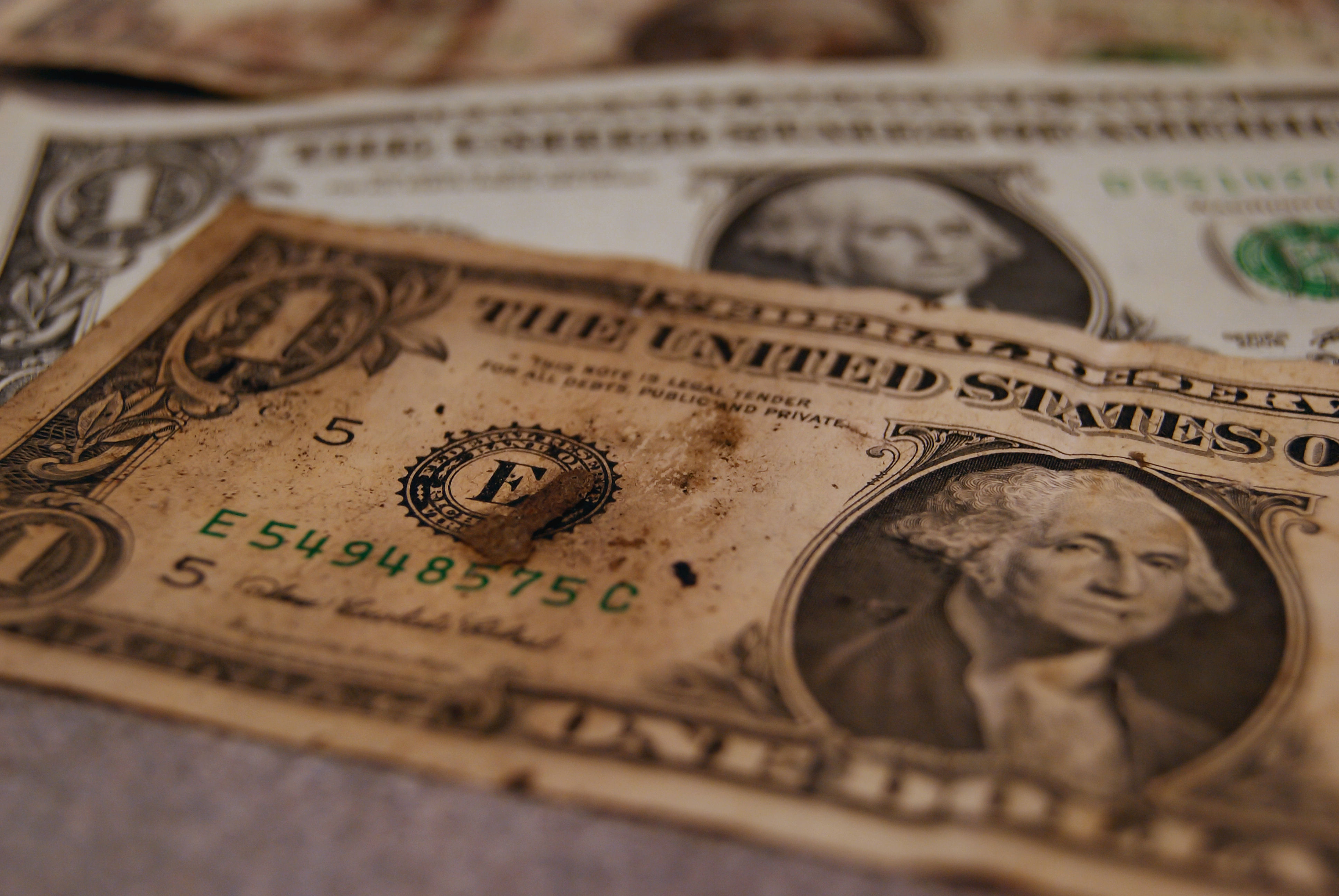
The phrase “Dirty money” may have different meanings for different people…but as far as microbiology is concerned….have you ever thought about what else you are getting back along with your change when you’re doing your shopping? I’m sure most of us are more concerned about getting the correct change rather than what microbes come for free with our change. For the few who have thought about what else they may be getting, I suspect that even fewer would answer: “pathogenic and sometimes multidrug-resistant bacteria, fungi and human parasites.”

In reality, we shouldn’t be surprised that bank notes and coins are contaminated with various bacteria. After all, we hardly expect them to be sterile. Our own hands are colonized with millions of bacteria and money is the most frequently passed item in the world. All studies that I have come across investigating bacterial contamination on money (paper notes or coins) have found a significant proportion to be contaminated (53-100%). I suspect that the microbiological techniques used in the various studies impacted on the results and my personal view is that the majority if not all money commonly exchanged between people will be contaminated.

The level of contamination and type or organisms on the money vary depending on the country, season, environmental conditions, type of money (paper vs coins), the type of material the money is made off, local community flora, the general hygiene level of the population and who is likely to be handling the money. Also dirty/damaged money (indication of frequent exchange) has been shown to be significantly more contaminated than clean and mint condition currency notes, and low denomination notes were more likely to be contaminated than higher denomination notes (probably reflecting frequency of use and socio-economic factors).1

The question to be asked, given the above, is: does it matter if money is contaminated with organisms? After all, this is not a new problem, as early as 1949, Nisbet and Skeoch2 have highlighted this issue. To answer this we need to look at a number of factors. These include: what type of organisms are on the money, how long are they able to survive and are they able to be transmitted to people and throughout the community from these contaminated currencies.  So let’s look at these factors in more detail:

1-  The type of organisms found on money

It is expected that bacteria will be found on bank notes and coins regardless of which country the money is from. Studies from Mexico, USA, India, Saudi Arabia, Nigeria, Kenya, Burma, China and Turkey to name few have all found significant contamination on their money. The type or bacteria found on money includes [deep breath]: E. coli, Vibrio spp., Klebsiella spp. including K. pneumoniae, Serratia spp., Enterobacter sp., Salmonella spp., Acinetobacter spp., Enterococcus spp., Staphylococcus including S. aureus, Bacillus spp., Staphylococcus epidermidis, Streptococcus pneumoniae, Proteus spp., Pseudomonas spp. including P. aeruginosa, Shigella spp.,  Corynebacterium, Lactobacillus spp., Burkholderia cepacia, Micrococcus spp. and Alcaligenes.

Looking at this list, it is clear that some of these bacteria are common environmental bacteria considered non-pathogenic. However, many are either potentially pathogenic or common human pathogens. For example, K. pneumoniae is a virulent organism and may cause both community and hospital-acquired infections. Even those organisms not commonly associated with disease in healthy hosts can cause clinically significant infections in immuno-compromised and hospitalised patients. These include even the natural inhabitants of the human skin such as Staphylococcus spp.

The story doesn’t end there since a number of studies have found multidrug-resistant and virulent strains on money. These have the potential to cause serious infections that are hard to treat, to disseminate in healthcare and community settings, and to spread antimicrobial resistant determinants to other bacteria. For example, one study3 found substantial S. aureus colonies on all 8 of the $1 and $5 bank notes collected from and around a University hospital in the USA. Tests for the presence of β-lactamases were positive and a significant number of the colonies showed resistance to erythromycin, tetracycline, chloramphenicol and vancomycin. Two isolates showed high-level resistance to vancomycin were found to harbour a plasmid conferring resistance to the drug. Taking that vancomycin is one of the last line antibiotics for treating multidrug resistant infection, this finding is very alarming. [A cautionary aside though – this work was published in the ‘Journal of Young Investigators’, so I can’t help thinking that the high-level resistance to vancomycin warrants some further investigation.] In another study,4 virulence genes were detected in S. aureus isolated from paper currency in India. Four virulence genes (cna, icaA, hlg and sdrE) were found in the isolates with 8 isolated possessing all 4 genes. Isolates harbouring these virulence genes showed higher antimicrobial resistance than those which didn’t contain these genes.

Bacteria are not the only organisms found on money. A number of studies show that fungal contamination of money is also common. Some of these are potentially pathogenic to humans and other life forms including plants. This may have implications far beyond human health to economic consequences if non-native pathogenic species are introduced into different countries via money carried during travel. For example, studies have found Penicillium spp., Aspergillus niger and A. flavus, Candida spp., Fusarium spp., Rhizopus spp., Alternaria spp, Trichoderma virie and white and brown mycelium on money.5.6 Some of these fungi can cause serious infections in humans and diseases in plants. In some countries, even parasites have been identified on bank notes. One study1 from Nigeria found that of the 250 currency notes collected from 4 major cities in the country, 21.6% were contaminated with enteric parasites including Ascaris lumbricoides, Enterobius vermicularis, Trichuris trichiura and Taenia spp. This parasite-contaminated currency was most frequently found in notes obtained from butchers and beggars. In another study,7 60.2% of 103 banknotes and 56.6% of 99 coins obtained from food-related workers in Egypt were found to be contaminated with one or more parasitic species. Protozoa were the predominant parasites, with microsporidia and Cryptosporidium spp. being the most prevalent.

2-  How long are organisms able to survive on money?

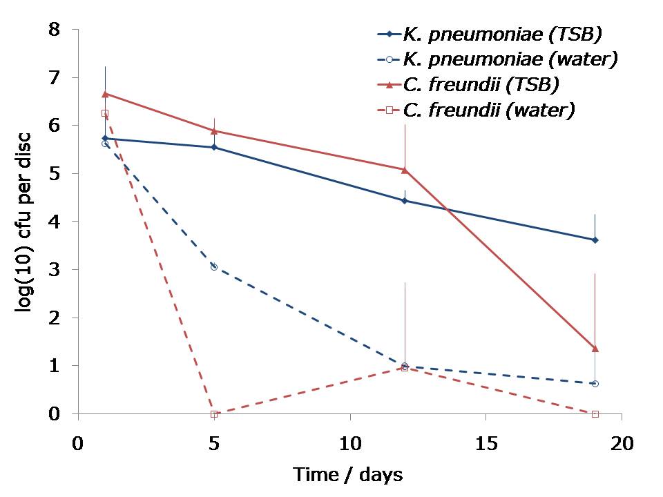
The survival of organisms on money depends on the type of the organism and their environmental resilience, the environmental conditions and the type of material the money is made of. Banknote paper is manufactured from cotton fibre, which gives the paper its strength and durability. Other additional elements maybe added to the cotton. Ploymer (or plastic) bank notes were developed to improve durability and incorporate some security features. One study8 investigated survival of MRSA, VRE and ESBL-producing E. coli on various bank notes from around the world including Euro, Croatian Kuna, Romanian Leu, Moroccan Dirham, US Dollar, Canadian Dollar, and the Indian Rupee. They found that the 3 organisms survived on the Romanian Leu for 6 hours after drying and VRE was isolated from the same notes after one day of drying. Other currencies had variable survival rates. Another in-vitro study4 found that S. aureus was able to survive on Indian paper currency for 8 days at room temperature.

3-  Are organisms able to be transmitted from money?

Transmission of organisms from money is highly significant if it occurs. For example, transmission from the community to the hospital setting is relevant because normally non-pathogenic, or opportunistic pathogens can have a serious clinical impact in such settings. On the other hand, transmission from the healthcare environment to the community is relevant when antimicrobial resistant strains (commonly found in hospitals) are involved. A study mentioned above,3 found vancomycin-resistant S. aureus on bank notes. The resistant determinant was located on a plasmid, hence easily transferrable. Notwithstanding my reservations about this study (see above), the interesting point about this investigation was the sources of the bank notes tested. These were collected from a University Hospital’s gift shop, a snack cart outside the hospital’s door and a convenience store near the hospital. The vancomycin-resistant isolates have likely originated from the hospital where the antibiotic is commonly used and had been transmitted to outside the hospital on money. In another study,8 investigators artificially contaminated bank notes of a number of countries with S. aureus and E. coli, and investigated transmission after 3 subjects with disinfected hands came into contact with these notes. Transmission was not successful for the Euro notes but transmission from US Dollars and the Romanian Leu was observed.

So we probably should be concerned with contamination of money especially when virulent, pathogenic or multidrug-resistant strains are concerned. Transmission between the healthcare and community settings can also have important implications. What’s the solution? Disinfection of the currencies in banks with UV light, supersonic or chemical means, producing bank notes from materials which inhibit bacterial growth or material with antimicrobial activity as well as replacement of traditional methods of trading with electronic money transactions,  have all been proposed. Personally I think for now, proper hand hygiene and overall hygiene remain the best ways to counter this problem.

References

  1. Uneke CJ, Ogbu O. Potential for parasite and bacteria transmission by paper currency in Nigeria. J Environ Health. 2007;69:54-60.
  2. Nisbet BR, Skeoch T. Bacteria on bank notes. Med Off. 1949;81:225.
  3. Bhalakia N. Isolation and plasmid analysis of vancomycin-resistant Staphylococcus aureus. J Young Investigators. 2005.
  4. Kumar JD, Negi YK, Gaur A, Khanna D. Detection of virulence genes in Staphylococcus aureus isolated from paper currency. Int J Infect Dis. 2009;13:e450-5
  5. Wanule D, Jalander V, Gachande BD, Sirsikar AN. Currency notes and coins as a possible source of transmitting fungal pathogens of man and plants. J Environ Sci Eng. 201;53:515-8.
  6. Kuria JK, Wahome RG, Jobalamin M, Kariuki SM. Profile of bacteria and fungi on money coins. East Afr Med J. 2009;86:151-5.
  7. Hassan A, Farouk H, Hassanein F, Abdul-Ghani R. Currency as a potential environmental vehicle for transmitting parasites among food-related workers in Alexandria, Egypt. Trans R Soc Trop Med Hyg. 201;105:519-24.
  8. Gedik H, Voss TA, Voss A. Money and transmission of bacteria. Antimicrob Resist Infect Control. 2013;2:22.

Photo credit: Sam Setzler.